Preclinical evaluation of 68Ga-labelled Pt(IV)-DFO complexes as PET-imaging agents for osteosarcoma
2026.09.03.
López-Martínez et al., European Journal of Medicinal Chemistry, 2026
Summary
The most commonly used chemotherapy agents for osteosarcoma are platinum(II)-based compounds; however, platinum(IV) prodrugs have recently gained attention as alternatives designed to overcome the dose-limiting toxicities and resistance mechanisms associated with Pt(II) complexes. In this study, Pt(IV) prodrugs were labeled with gallium-68 68Ga using deferoxamine (DFO) as a chelator to facilitate in vivo tracking via positron emission tomography (PET) imaging. Five complexes were evaluated: four functionalized Pt(IV)-DFO complexes (A–D) and one DFO-succinic acid conjugate (1). Comprehensive in vivo characterization of the PET tracers was conducted in both naïve and orthotopic osteosarcoma mouse models. Prior to administering the 68Ga-labeled Pt(IV)-DFO tracers, tumor-bearing mice underwent 18F-FDG PET scans to baseline and monitor tumor growth
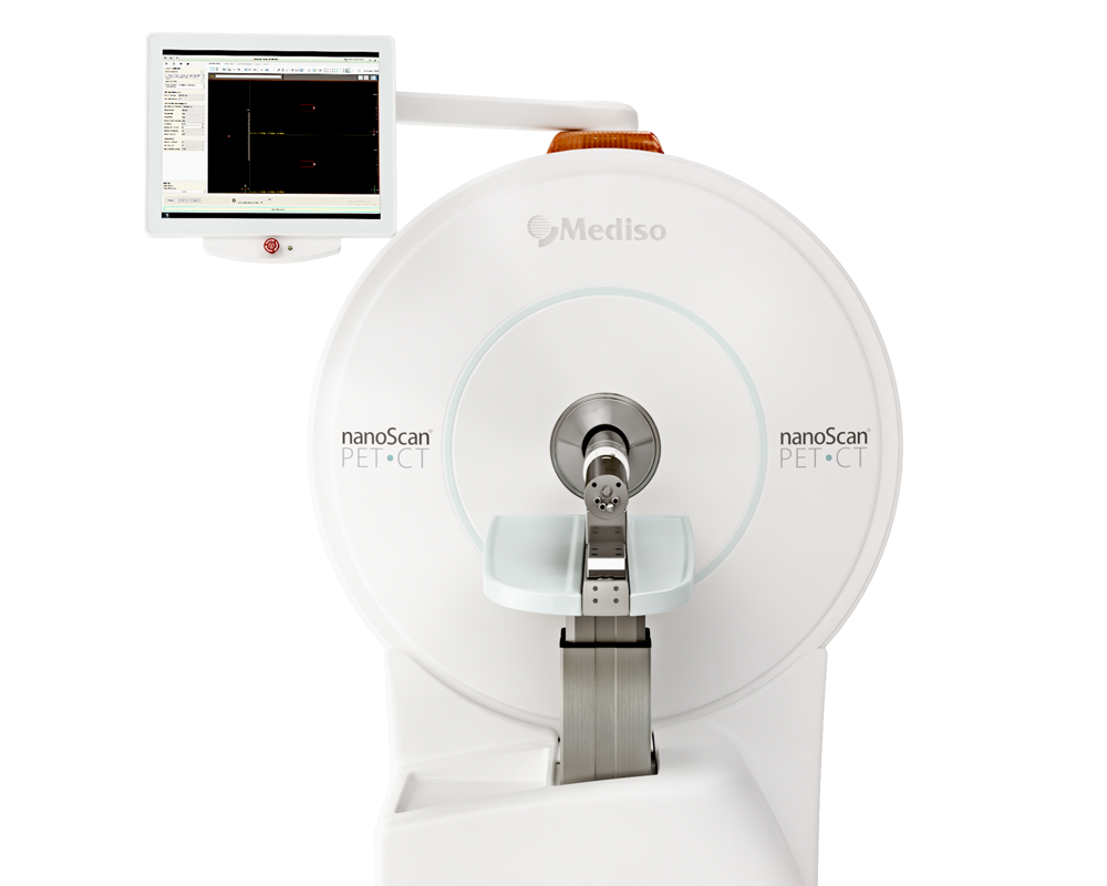
Results from nanoScan® PET/CT
Both naive (n = 12; 6 female/6 male per tracer) and osteosarcoma model mice (n = 6 per tracer) underwent dynamic PET imaging. Anesthesia was induced using isoflurane, and a 30-gauge tail-vein catheter was inserted for tracer administration. Respiration was continuously monitored, and body temperature was maintained using a heating pad set to 38 °C throughout the scan. Two animals were imaged simultaneously using a dual-bed setup per imaging session. For the 68Ga-labeled Pt(IV)-DFO measurements, 11–15 MBq in 100 μL was injected, followed by a 60-minute dynamic PET acquisition and subsequent CT imaging (720 projections, 50 kV, 980 μA, 300 ms exposure time). Tumor progression was monitored on days 7, 10, and 14 post-inoculation using 18F-FDG. For these scans, a 10-minute static PET acquisition was initiated 60 minutes post-injection, followed by low-dose CT imaging (360 projections, 50 kV, 980 μA, 170 ms exposure time).
Raw PET data were reconstructed using the Tera-TOMOTM iterative reconstruction algorithm (8 subsets, 4 iterations) with an isotropic voxel size of 0.4 mm3. Dynamic PET data were frame-binned into time windows 8 × 5 s, 2 × 10 s, 2 × 30 s, 3 × 60 s, 2 × 150 s, 2 × 300 s. CT projections were reconstructed using filtered back-projection (FBP), resulting in isotropic voxel sizes of 125 µm3 for standard CT scans and 250 µm3 for low-dose CT scans.

Fig. 1 (I) In vivo PET/CT images of female naïve mice at 15 min post-injection from all 68Ga-labelled compounds. Coronal sections show predominately renal uptake (A-E), and intestinal/hepatic distribution (F-J). The white arrows indicate different organs of interest, K: kidney; B: bladder; SI: small intestine; L: liver. (II) In vivo PET/CT images of osteosarcoma-bearing mice at 12.5 min post-injection of all different 68Ga-labelled compounds. The white arrow indicates the tumour region (T) in the left hind limb in the first image; the tumour remains in the same anatomical position across all subsequent images. (III) Area under the curve (AUC0-55min) derived from PET time-activity curved for tumour and healthy leg tissues across all 68Ga-labelled compounds. Statistical analyses were performed using a paired t-test (p < 0.05 (*), p < 0.01 (**)).
The general conclusion of this study is that the 68Ga-labeled Pt(IV)-DFO complexes performed remarkably well in the orthotopic osteosarcoma model, with [68Ga] Ga-D demonstrating the most favorable kinetic profile. Although further preclinical evaluation is required, these tracers hold significant potential for future clinical translation.
Full article on sciencedirect.com
How can we help you?
Don't hesitate to contact us for technical information or to find out more about our products and services.
Get in touch