Panitumumab labeled with Auger electron-emitting 197gHg/197mHg: cytotoxicity on human breast cancer cells and tumour and normal tissue uptake in mice with EGFR-overexpressing breast cancer xenografts
2026.07.13.
Arthur C. K. Chu et al., EJNMMI Radiopharmacy and Chemistry, 2026
Summary
The objective of this study was to evaluate whether the Auger electron-emitting radionuclides ¹⁹⁷ᵍHg/¹⁹⁷ᵐHg, attached to the anti-EGFR antibody panitumumab, could serve as a theranostic agent for EGFR-positive triple-negative breast cancer (TNBC). The authors aimed to
- Develop methods for labeling panitumumab with ¹⁹⁷ᵍHg/¹⁹⁷ᵐHg.
- Determine whether the radiolabeled antibody specifically binds to EGFR-positive breast cancer cells.
- Assess cellular uptake, nuclear localization, DNA damage, and cytotoxicity in vitro.
- Evaluate tumor targeting and normal tissue uptake in mice using SPECT/CT imaging and biodistribution studies
The study demonstrated that ¹⁹⁷ᵍHg/¹⁹⁷ᵐHg-labeled panitumumab specifically targets EGFR-expressing breast cancer cells, is internalized into the nucleus, induces DNA double-strand breaks through Auger electron emission, and markedly reduces clonogenic survival of EGFR-high tumor cells.
Results from nanoScan® SPECT/CT
In vivo SPECT/CT confirmed tumor targeting; however, substantial kidney uptake indicated partial release of mercury from the antibody in vivo. The authors conclude that the approach shows promise for theranostic imaging and Auger electron radioimmunotherapy, but improved radiolabeling methods are needed to increase in vivo stability and reduce renal accumulation.
- visualization of EGFR-positive tumors in mice following administration of radiolabeled panitumumab
- Tumor uptake was visible on both 1-day and 3-day SPECT/CT images
- Tumor uptake decreased when excess unlabeled panitumumab was co-administered, demonstrating EGFR-specific targeting
- Free ¹⁹⁷HgCl₂ showed minimal tumor uptake, confirming that antibody targeting was responsible for tumor localization
- High renal uptake was observed for all radiolabeled antibodies
- Biodistribution measurements confirmed the SPECT/CT imaging findings
- Imaging demonstrated that all panitumumab constructs localized to tumors, although in vivo instability likely resulted in partial mercury release and kidney accumulation
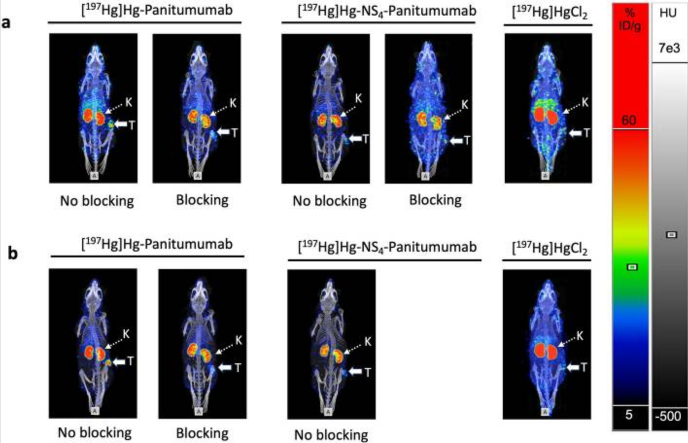
Figure 7. Representative SPECT/CT images of CD1 athymic (nude) mice bearing subcutaneous MDA-MB-468 human EGFR-overexpressing breast cancer xenografts following intravenous administration of Hg-Panitumumab , Hg-NS4-Panitumumab, or HgCl₂. Mice received each tracer either alone (No blocking) or together with a 10–30-fold excess of unlabeled panitumumab (Blocking) to competitively inhibit EGFR binding. (a) Images acquired 1 day post injection (p.i.). (b) Images acquired 3 days p.i. Tumours (T, solid arrows) are visualized with both antibody tracers, while EGFR blocking reduces tumour uptake, particularly at 3 d p.i., indicating receptor-specific targeting. HgCl₂ showed no detectable tumour uptake at either time point. Kidneys (K, dashed arrows) exhibited high uptake for all tracers, with the highest renal accumulation observed for HgCl₂. Color scale represents %ID/g overlaid on CT images (HU).
How can we help you?
Don't hesitate to contact us for technical information or to find out more about our products and services.
Get in touch