In Vitro and Preclinical Systematic Dose-Effect Studies of Auger Electron- and β Particle-Emitting Radionuclides and External Beam Radiation for Cancer Treatment
2024.05.24.
Ines M. Costa et al., International Journal of Radiation Oncology, Biology, Physics, 2024
Summary
Therapeutic radiopharmaceuticals deliver radioactivity to the target of interest in both primary and disseminated disease. Some radionuclides investigated for molecular radiotherapy (MRT) can be considered theranostic because they emit γ-rays or positrons (β+), which can be exploited for single-photon emission computed tomography (SPECT) or positron emission tomography (PET) imaging, respectively.
The field of MRT has been driven by β–-particle-emitters, such as ([177Lu]Lu-prostate-specific membrane antigen-617 [177Lu]Lu-PSMA-617 for patients with advanced prostate cancer. Current MRT research builds on previous successes with radiopharmaceutical therapies including, [131I]I-Meta-Iodo-Benzyl-Guanidine ([131I]I-MIBG) for neuroblastoma in children, [90Y]Y-anti-CD20 antibodies for non-Hodgkin lymphoma, [90Y]Y-microspheres for hepatic tumors, and 131I for thyroid cancer. The success of MRT has largely been energized through its implementation in patients with advanced midgut neuroendocrine tumors and [Lu]Lu-[DOTA0-Tyr3]-octreotate ([177Lu]Lu-DOTA-TATE) is now a well-established therapeutic option. Brachytherapy using β−-emitting [188Re]Re-microspheres also has clinical potential in patients with hepatocellular carcinoma. Studies also suggest that 188Re could become a key isotope in targeted MRT, for example, as [188Re]Re-anti-CD20 for non-Hodgkin lymphoma or other targeted deemed useful for MRT.
The MRT radioisotope portfolio is currently expanding to include radionuclides that emit α-particles, beyond 223Ra, to Auger electron (AE)–emitting radionuclides. AE radiation therapy exploits the cytotoxicity of low-energy electrons emitted during radioactive decay that travel very short distances (typically <1 μm, 4-26 keV/μm). For example, 99mTc(I) tricarbonyl complexes containing a triphenylphosphonium and bombesin peptide have proved effective at killing prostate cancer cells in vitro as did [99mTc]Tc-labeled doxorubicin in HeLa, melanoma B16, and epidermoid carcinoma A-431 cancer cell lines. The potential of AE radiation therapy is also exemplified by interest in developing clinical dose escalation trials, for example, for [111In]In-diethylenetriamine pentaacetate-human epidermal growth factor ([111In]In-DTPA-hEGF) in patients with metastatic breast cancer, and advances toward treating patients with glioblastoma with an [123I]I-labeled poly (ADP-ribose) polymerase 1 (PARP1) inhibitor.
Currently, there are few biological studies directly comparing the radiobiological effects of different radionuclides in the same model; most work in this area used bacterial plasmids. These assays do not account for any complex cellular environment or longer-distance effects, for example, crossfire effect. As such, few studies correlate radiobiological effects with the delivered radiation absorbed dose at the cellular level. This knowledge is key to reach a better understanding of the consequences of ionizing radiation exposure in biological matter, and ultimately boost radiopharmaceutical development for more effective MRT strategies for cancer treatment.
Here, the authors employed triple-negative breast cancer cells, previously engineered to express the human sodium iodide symporter (hNIS) in which [99mTc]TcO4- proved an effective radiotherapeutic. In this work, the same approach was used in vitro and in vivo to determine and compare the biological effect of 2 additional sodium iodide symporter substrates, [123I]I− and [188Re]ReO4−, alongside X-ray radiation (external beam radiation therapy [EBRT]) as a comparator for which the biological effects relating to radiation absorbed dose are well understood.
Results from the nanoScan SPECT/CT
Radionuclides were delivered to mice as described above, and SPECT/CT scans were acquired over a period of 1.5 hours and at 5 hours and 24 hours after radiotracer administration.
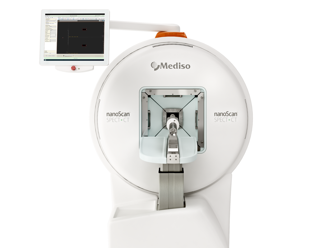
For imaging, anaesthetized mice were placed in prone position on the warmed (37○C) nanoSPECT/CT scanner bed (Mediso, Hungary), with anaesthesia maintained until the end of each imaging session. Radionuclides were delivered as described above. SPECT scans were acquired over a period of 1h 30 min (6 x 15 min sequential SPECT scans, 15 sec per frame, 360° rotation, low energy multi-pinhole with 1 mm aperture and 9 holes and 20% energy window centred on photo-peaks of 123I (159 keV: 143-175 keV) and 188Re (155 keV: 140-171 keV). At the end of the sequential SPECT scans, a single 20 min CT scan (55 kVp X-ray, exposure time 1000 ms, 360○ rotation and pitch 1 was performed. Mice were then allowed to completely recover from anaesthesia, and then re-anaesthetized and re-scanned by SPECT/CT at 5 hours and 24 hours after radiotracer administration. At 5 hours post administration, mice underwent a 45-minute SPECT scan (same acquisition parameters, but with 60 seconds per frame) followed by a 20-minute CT scan. At 24 hours post administration, mice underwent a 2-hour SPECT scan (same acquisition parameters, but 150 sec per frame) followed by a 20-minute CT scan.
All SPECT projections were reconstructed using Tera-TomoTM iterative algorithm (Mediso, Hungary; reconstruction (64 iterations, high regularization filter) with attenuation and scatter correction. Data was visualized and quantified using VivoQuant© v3.5-patch2 software (Invicro, Massachusetts, USA). Volumes of interest were manually delineated on the stomach, thyroid, salivary glands, lacrimal glands, hNIS-expressing tumors, kidney, bladder, and muscle. CT scans were used as an anatomic reference to define the boundary of the organs and tumors. SPECT/CT images were shown as maximum intensity projections (MIPs) and data was expressed as percentage of injected activity per mL (%IA/mL) using GraphPad Prism 9.1.0. All images were decay-corrected to the time of radiotracer administration.
After the imaging protocol detailed above, mice were longitudinally monitored, and tumor volume and body weight were measured.
Fig. 3A. shows maximum intensity projections of single-photon emission computed tomography/computed tomography images of [123I]I− in percentage activity per milliliter (%IA/mL) up to 24 hours after administration of 55 MBq [123I]I−. Image-based time-activity curves: %IA/mL up to 24 hours after administration of (B) [123I]I− and (C) [188Re]ReO4− in hNIS-GFP–expressing tumors and NIS-expressing organs. Time-activity curves were decay-corrected to time of radionuclide administration. N=4 to 6 mice/group. Abbreviations: LG = lacrimal glands; S = stomach; T = MDA-MB-231.hNIS-GFP tumors; Th = thyroid.
Fig. E11. shows SPECT/CT image-based biodistribution of [188Re]ReO4─ up to 24 hours post intravenous administration of 55 MBq. Maximum intensity projections (MIPs) show the percentage activity per mL (% IA/mL) of radionuclides in endogenously NIS-expressing organs (white inscriptions: LG-lacrimal glands, Th- thyroid, and S-stomach), MDA-MB-231.hNIS-GFP tumors (orange inscription: T- tumor). CT images (grayscale) were used as anatomical reference.
- This work showcases the power of using hNIS models to permit systematic comparative radionuclide investigations, here exemplified with [99mTc]TcO4−, [123I]I−, and [188Re]ReO4−.
- The authors also report reference data, for the first time, compared in cell and tumor models that were fully balanced for all radionuclides used.
- They further demonstrate the relative tumor-controlling effects of [123I]I−, [188Re]ReO4−, and EBRT, whereby we found MRT with [188Re]ReO4− to be able to counteract tumor dissemination in this model.
How can we help you?
Don't hesitate to contact us for technical information or to find out more about our products and services.
Get in touch
