HER2 expression in different cell lines at different inoculation sites assessed by [52Mn]Mn-DOTAGA(anhydride)-trastuzumab
2025.04.29.
Toàn Minh Ngô et al., Pathol Oncol Res., 2025
Purpose
Positron emission tomography (PET) hybrid imaging targeting HER2 requires antibodies labelled with longer half-life isotopes. With a suitable radiation profile, 52Mn coupled with DOTAGA as a bifunctional chelator is a potential candidate. In this study, the authors investigated the tumor HER2 specificity and the temporal biodistribution of the [52Mn]Mn-DOTAGA(anhydride)-trastuzumab in preclinical models.
Methods
PET/MRI and PET/CT were performed on SCID mice bearing orthotopic and ectopic HER2-positive and ectopic HER2-negative tumors at 4, 24, 48, 72, and 120 h post-injection with [52Mn]Mn-DOTAGA(anhydride)-trastuzumab. Melanoma xenografts were included for comparison of specificity.
Results
In vivo biodistribution demonstrated strong contrast in HER2-positive tumors, particularly in orthotopic tumors, where uptake was significantly higher than in the blood pool and other organs from 24 h onwards and consistently higher than in ectopic HER2-positive tumors at all time points. Significantly higher tumor-to-blood and tumor-to-muscle ratios were observed in HER2-positive ectopic tumors compared to HER2-negative tumors but only at 4 and 24 h; the differences were likely due to non-specific binding of the tracer. The ratios for orthotopic HER2-positive tumors were significantly higher than those for ectopic HER2-negative tumors and melanoma at all time points. However, the differences between HER2-positive and HER2-negative tumors decreased at later time points.
Results from nanoScan® PET/MRI and nanoScan® PET/CT
52Mn is a potential candidate for antibody-based imaging, offers a superior radiation profile compared to 89Zr and 64Cu. With a T1/2 = 5.6 days and a 29% beta-positive emission with low maximum energy (0.575 MeV), 52Mn provides a short-tissue penetration and an improved resolution to be used for PET scans.
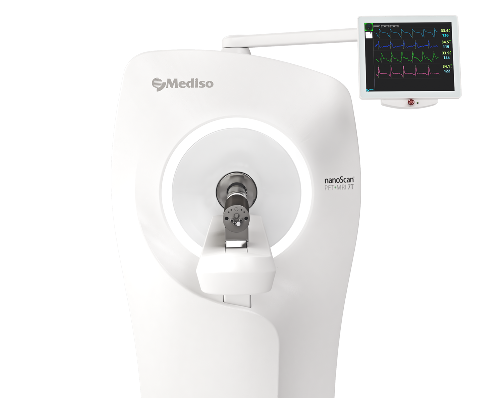
In the current study all mice were injected with 3.50 ± 0.59 MBq of [52Mn]Mn-DOTAGA-trastuzumab intravenously into the lateral tail veins. The HER2-positive groups were scanned with hybrid cameras nanoScan® PET/CT (Mediso Ltd., Hungary), while the HER2-negative and melanoma-bearing mice underwent scans with nanoScan® PET/MRI 1T (Mediso Ltd., Hungary). The scans were conducted using a scanning bed to minimize model movements, while closely monitoring temperature, heart rate, and respiratory rate throughout the procedure. The PET static scans with a duration of 20 min each scan was performed at 4 h, 24 h, 48 h, 72 h, and 120 h post-injection. The anatomical images for localization and attenuation correction maps were obtained by either MRI T1 gradient echo with 0.5 mm slice thickness, 20 ms repetition time, 2.6 ms echo time, and 20° flip angle or CT with 180 projections and 55 kVp X-ray source. The image reconstruction was done by Nucline software (Mediso Ltd., Hungary) using the Tera-Tomo™ 3D maximum-likelihood expectation-maximization (MLEM) method with attenuation correction, random correction, and scatter correction. Using the InterView™ FUSION software (Mediso Ltd., Hungary), volumes of interest (VOIs) and regions of interest (ROIs) were delineated on the reconstructed images.
Results show:
- By using the nanoScan® PET scanner it was possible to visualize [52Mn]Mn-DOTAGA-trastuzumab tracer which is an attractive but underexplored option for antibody labelling, with only a few studies investigating its potential. Additionally, the paramagnetic properties of manganese (II) make it a valuable tool for MRI studies, especially in the expanding field of hybrid cameras.
- On the PET images the orthotopic tumors consistently exhibited significantly greater tracer activity than the ectopic tumors at all time points. This difference increased over time and peaked around day 3, with breast tumor. Similarly, the orthotopic tumors showed significantly higher tumor-to-background contrast, becoming clearly visible from the first time point, whereas the ectopic tumors showed lower contrast, with only peripheral areas being highlighted at most time points. See figure 3.

Figure 3. Images of PET/MRI and PET/CT scans were obtained at different time points (4, 24, 48, 72, 120 h) following injection of the tracer into MDA-MB-468 tumour-bearing mice (upper row) and into the MDA-MB-HER2+ tumour-bearing mice (middle and bottom rows displaying the back and breast tumours, respectively). The upper row displays sagittal planes, with the exception of the 48-hour image in coronal plane. The middle and bottom rows display coronal planes. Red circles were used to highlight the MDA-MB-468 back tumour (upper row), the MDA-MB-HER2+ back tumour (middle row), and the MDA-MB-HER2+ breast tumour (bottom row) at 4 h and 120 h time points.
-
When comparing the tumor-to-background ratios of the xenografts visualized by the PET scans, the ectopic HER2-positive tumors showed significantly higher ratios than the HER2-negative tumors at 4 h post-injection; however, these differences began to decrease from 24 h onwards. Significantly higher ectopic HER2-positive tumor-to-blood ratios than ectopic melanoma ratios were observed from 24 h, but the difference was not significant using tumor-to-muscle-ratios.
-
Low urinary bladder activity was demonstrated by subsequent PET scans that indicates stable conjugation of the tracer in vivo. The complexation of the tracer seemed to be stable in vivo as well, as suggested by the minimal uptake in the pancreas, salivary glands, and joints without renal reabsorption.
The authors applied tumor-to-background ratio for assessment, aiming to ensure accurate and reliable results. Using the ratios, such as SUVmean Organ/Muscle, provided reproducible results, as demonstrated by the comparable biodistribution observed in the two breast cancer-bearing groups scanned with different modalities.
Conclusion
This study demonstrated high tumor/non-tumor ratios and the relative stability of [52Mn]Mn-DOTAGA(anhydride)-trastuzumab and highlighted the influence of inoculation sites, tumor characteristics, and the microenvironment on tumor tracer uptake. Despite its ease of production, the tracer exhibits noticeable non-specific binding and requires further improvement in immunoreactivity. Nevertheless, 52Mn-labelled trastuzumab has great potential for in vivo HER2 assessment as it allows non-invasive longitudinal characterization of HER2 expression in tumors and whole-body imaging to detect unusual tracer accumulation.
Full article on por-journal.com
How can we help you?
Don't hesitate to contact us for technical information or to find out more about our products and services.
Get in touch
