Performance evaluation of a novel multi-pinhole collimator for dopamine transporter SPECT
2020.08.14.
K Tecklenburg et al., Physics in Medicine & Biology, 2020
Abstract
There is a tradeoff between spatial resolution and count sensitivity in SPECT with conventional collimators. Multi-pinhole (MPH) collimator technology has potential for concurrent improvement of resolution and sensitivity in clinical SPECT of ‘small’ organs. This study evaluated a novel MPH collimator specifically designed for dopamine transporter (DAT) SPECT with a triple-head SPECT camera. Count sensitivity was measured with a 99mTc point source placed on the lattice points of a 1 cm grid covering the whole field-of-view (FOV). Spatial resolution was assessed with a Derenzo type hot rod phantom. An anthropomorphic striatum phantom was scanned with total activity representative of a typical patient scan and different striatum-to-background activity concentration ratios. Recovery of striatum-to-background contrast was assessed by the contrast-recovery-coefficient. Measurements were repeated with double-head SPECT with fan-beam or low-energy-high-resolution-high-sensitivity (LEHRHS) collimators. A patient referred to DAT SPECT because of suspicion of Parkinson’s disease was scanned with both LEHRHS and MPH collimators after a single tracer injection. The axial MPH sensitivity profile was approximately symmetrical around its peak, although it was shifted 7 cm towards the patient to simplify positioning. Peak sensitivity of the triple-head MPH system in the center of the FOV was 620 cps MBq−1 compared to 225 cps MBq−1 for the double-head fan-beam system. Sensitivity of the MPH system decreased towards the edges of the FOV. The full width of the sensitivity profile at 200 cps MBq−1 was 21 cm transaxially and 11 cm axially. In MPH SPECT of the Derenzo phantom all rods with ≥ 5 mm diameter were clearly visible. MPH SPECT improved striatal contrast recovery by ≥ 20% compared to fan-beam SPECT. The patient scan demonstrated good image quality of MPH SPECT with almost PET-like delineation of putamen and caudate nucleus. SPECT with dedicated MPH collimators provides considerable improvement of the resolution-sensitivity tradeoff in DAT SPECT compared to SPECT with fan-beam or LEHRHS collimators.
Methods
Triple-head SPECT system
The AnyScan® TRIO (Mediso Medical Imaging Systems, Budapest, Hungary) is a general purpose triple-head SPECT system. Each head is equipped with a 3/8” NaI(Tl) detector of 585 mm (transaxial) x 470 mm (axial), 16 mm lightguide and 94 photomultipliers. MPH collimators specifically designed for DAT SPECT are available for this system in addition to conventional parallel-hole collimators.
Multi-pinhole collimator and Tera-TomoTM reconstruction
The MPH collimator (figure 1) was designed for targeted SPECT imaging with high count sensitivity at the striatum and minimum multiplexing artefacts by combination of overlapping and non-overlapping projections. The collimator features a solid tungsten aperture plate of 18 mm thickness with 20 pinholes approximately arranged in 5 axially oriented columns and 4 transaxially oriented rows (figure 1(C)). The MPH was designed for a total SPECT field-of-view (FOV) of 220 mm transaxial diameter and 180 mm axial length (figure 1(A)).

Figure 1. (A) Drawing of the detector heads equipped with the multi-pinhole collimator (only two of the three detector heads are shown). The pinhole axes are tilted towards the patient such that the field-of-view (FOV) is shifted 7 cm towards the patient in axial direction. This simplifies positioning the striatum in the central FOV (CFOV) also in patients with short neck. Part (B) shows a photograph of the rear view of the MPH collimator. Part (C) is a drawing showing the arrangement of the 20 pinholes on the tungsten aperture plate. Part (D) is a drawing of the projection pattern on the scintillation crystal to illustrate the combination of non-overlapping and overlapping projections. Part (E) shows the projection pattern of a 57Co flood phantom placed directly on the touch plate on top of the aperture plate.
The MPH reconstruction is built upon the iterative one-step-late maximum-a-posteriori expectation-maximization scheme. A Monte Carlo photon simulation engine is used for forward and backward projection. The forward projector is divided into two parts. The first part is a ‘direct’ projection considering photons directed towards the pinholes and accounting only for attenuation (no scatter) in the patient (based on segmented CT if available). The second part simulates only scatter in the patient, starting from the count density at the current step of the iterative reconstruction. The photons simulated for scatter are emitted isotropically, that is, uniformly in the full 4π solid angle. The Woodcock algorithm is used to track the photon path and to simulate Compton scattering. Attenuation of scattered photons is also taken into account. The aperture plate of the MPH collimator and the detector crystal are simulated similarly, considering both Compton scatter and photo electric effect. Variance reduction methods are used such that Compton scatter is limited by the detectable energy defined by the energy window of the acquisition. Only photons with a low weight are removed (bias is restored using ‘Russian roulette’), and photons are forced to be detected in the crystal. The intrinsic spatial resolution of the detector is modeled by Gaussian convolution based on calibration data measured during installation of the SPECT system. The backprojection in MPH image reconstruction is a geometric one (with Monte Carlo sampling), it does not match with the forward projection.
Regularization is achieved by filtering between iterations and the beta parameter of the total variation prior used in the maximum-a-posteriori scheme. Filtering between iterations comprised two independent procedures, bright-dot removal and 3-dimensional bilateral filtering. Bright-dot removal works as follows: for each voxel, the bright-dot filter compares the count density in the considered voxel with the count density in the 26 neighboring voxels in the 3x3x3 voxels cube centered at the considered voxel. If the current count density in the considered voxel is identified as an outlier, it is replaced by the median count density in the 26 neighboring voxels. The voxel is considered an outlier if the count density in 23 or more of the 26 neighboring voxels differs more than 10% from the count density in the considered voxel. Bright-dot removal stabilizes the iterative reconstruction particularly in cases with high statistical noise. Bilateral-filtering is a filtering technique for edge-preserving denoising of images including low-count SPECT. In contrast to conventional filtering, the weights of bilateral filters depend not only on the distance of voxels but also on radiometric features such as count density. The latter allows to better preserve edges. The quality of the Monte Carlo simulation, associated with the number of simulated photons, can be chosen as low (8 million simulated photons), medium (16 million) or high (32 million).
Results
Count sensitivity
Profiles of system count sensitivity throughout the FOV of the triple-head system with the MPH collimators and the double-head system with fan-beam and LEHRHS collimators are shown in figure 8. Peak system sensitivity of the triple-head SPECT with MPH collimators was 620 cps MBq−1 compared to 225 cps MBq−1 for the double-head fan-beam system and 190 cps MBq−1 for the double-head LEHRHS system. Sensitivity of the MPH system decreased towards the edges of the FOV. The full width of the sensitivity profile at 200 cps MBq−1 was 11 cm in axial direction and 21 cm in transaxial direction (at the axial peak). The impact of varying table displacement for helical scanning on the axial sensitivity profile of the triple-head SPECT system with MPH collimators is shown in figures 8(E) and (F). Peak count sensitivity of the triple-head system with the MPH collimators measured with 123I (rather than 99mTc) was 614 cps MBq−1 and 598 cps MBq−1 without and with 40 mm table displacement for helical scanning, respectively.

Figure 8. System count sensitivity profile of the triple-head SPECT system with the MPH collimators compared to the double-head SPECT systems with fan-beam and LEHRHS collimators in axial direction along the center of rotation axis (A) and in transaxial direction at the maximum of the axial sensitivity profile (C). Parts (B) and (D) show the FWHM of the reconstructed point source images across the FOV. MPH SPECT images were acquired and reconstructed with parameter settings optimized for DAT SPECT in clinical routine (nv = 90, td = 40, low Monte Carlo quality, 90 effective iterations, 3 subsets), fan-beam SPECT images were reconstructed with the ordered-subsets-expectation-maximization algorithm with resolution recovery implemented in the HybridRecon-Neurology tool of the Hermes SMART workstation v1.6 with parameter settings recommended for clinical DAT SPECT by Hermes. Part (E) shows peak system sensitivity (left axis) and FWHM of the reconstructed point source image (right axis) at peak location of the triple-head SPECT with MPH collimators for varying total table displacement during helical scanning. Part (F) shows complete axial profiles of system sensitivity for selected table displacements (td = 0, 40, 80 mm).
Spatial resolution
SPECT images of the Derenzo type hot rod phantom filled with 99mTc- or 123I-solution are shown in figures 9(A) and (B), respectively. All rods with ≥ 5 mm diameter were clearly visible, all rods with ≤ 3 mm diameter could not be detected. Some of the 4 mm rods were visible, but with low contrast. The simulated images of the anthropomorphic striatum phantom with varying spatial resolution are shown in figure 9(C). According to visual inspection, the simulated SPECT image with 5 mm spatial resolution shows best agreement with the measured MPH SPECT image (figure 9(B)) with respect to spatial resolution. The FWHM of the reconstructed point source images did not reveal relevant non-uniformity of spatial resolution of MPH SPECT across the central FOV (figures 8(B), (D)).

Figure 9. Transaxial images of the Derenzo type hot rod phantom filled with 99mTc (A) or 123 I (B). Acquisition and reconstruction were performed with the parameter settings optimized for clinical DAT SPECT. The image was obtained by summing 16 transaxial slices of 1.72 mm thickness through the rods (resulting in 27.5 mm of total thickness) in order to reduce statistical noise. (C): Central transversal image of 2 mm thickness through the striatum in MPH SPECT of the anthropomorphic striatum phantom with true 4:1 (left striatum) and 3:1 (right striatum) contrast acquired with a total number of 17.5 million counts. Acquisition and reconstruction were performed with the optimized settings for clinical DAT SPECT (nv = 90, td = 40 mm, low Monte Carlo quality, 90 effective iterations, 3 subsets). CT-based attenuation and scatter correction was performed during iterative reconstruction. (D): Simulated SPECT images of 2 mm thickness of the anthropomorphic striatum phantom with 4:1 and 3:1 striatum-to-background contrast with 3, 4, …, 8 mm spatial resolution (without statistical noise).
MPH vs LEHRHS collimator
Superior imaging characteristics of MPH DAT SPECT compared to fan-beam DAT SPECT was confirmed in a patient scan (figure 12). Image quality of the MPH DAT SPECT in the patient was almost PET-like, although acquisition time was shorter compared to fan-beam SPECT (30 min versus 40 min).

Figure 12. Normal DAT availability in a male patient (54 y) referred to DAT SPECT because of suspicion of PD. The patient was scanned twice after a single 123 I-FP-CIT injection (182 MBq). (A): 40 min scan in double-head mode with the triple-head system equipped with LEHRHS collimators that was started 3 h 10 min after tracer injection. (B): 30 min scan with the triple-head system equipped with MPH collimators in triple-head mode that was started 5 h 7 min after tracer injection. Both images were reconstructed with the parameter settings optimized for DAT SPECT in clinical routine. The color indicates the distribution volume ratio (DVR) obtained by scaling the count density voxel by voxel to the 75-th percentile of the count density in a reference region comprising the whole brain except striata, ventricles, and thalamus.
Conclusion
MPH collimators provide considerable improvement of image quality in DAT SPECT with respect to both spatial resolution and statistical noise compared to conventional collimators including low-energy-high-resolution-high-sensitivity collimators and fan-beam collimators. As visual interpretation of DAT SPECT performed with conventional collimators by experienced readers already provides high diagnostic accuracy (≥ 90% sensitivity and specificity (O’Brien et al 2014)), we expect improved image quality in MPH DAT SPECT to be particularly useful in borderline cases and for less experienced readers. We also expect it to result in improved reader confidence independent of the reader’s experience. However, the largest benefit of improved image quality in DAT SPECT by MPH technology is expected for early detection (or exclusion) of PD before the loss of putaminal DAT reaches 50% and PD-characteristic motor symptoms occur. These hypotheses might be tested in prospective clinical studies, preferably by comparing MPH SPECT and parallel-hole/fan-beam SPECT acquired in randomized order after a single injection of FP-CIT in the same patients.
Full article on Physics in Medicine & Biology.
-
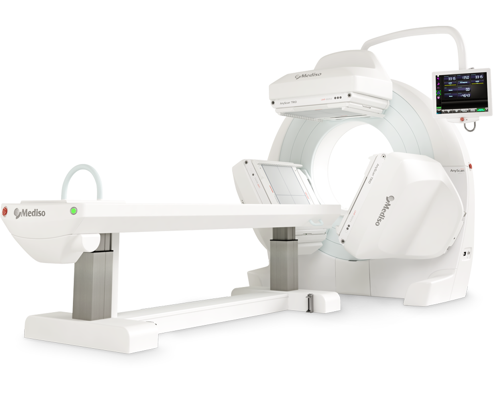
AnyScan® TRIO SPECT
Ultra-fast Triple-NaI-Detector SPECT for wide range of clinical applications
-
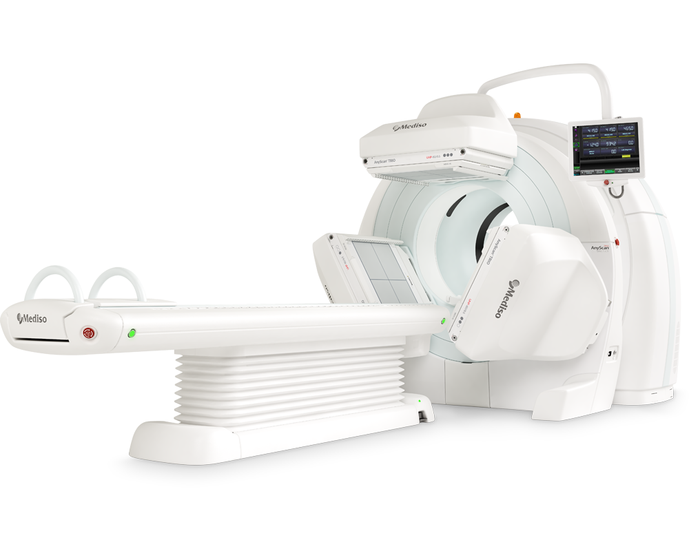
AnyScan® TRIO SPECT/CT
Ultra-fast Triple-NaI-Detector SPECT system for wide range of clinical applicati...
-
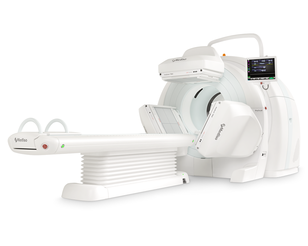
AnyScan® TRIO SPECT/CT/PET
An integrated triple-detector SPECT/CT and PET/CT system in a single-room instal...
-
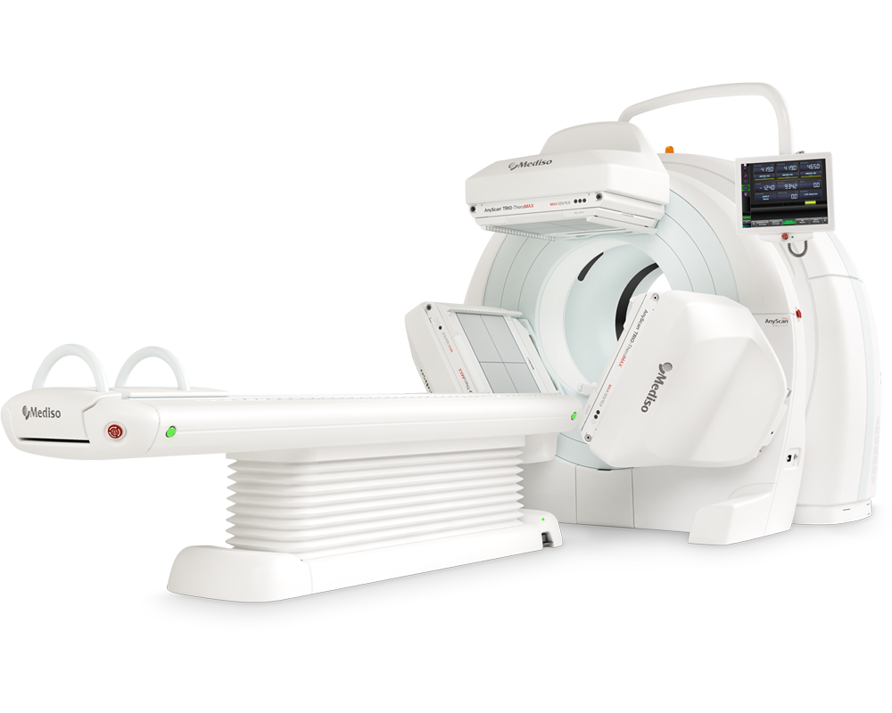
AnyScan® TRIO-TheraMAX SPECT/CT
Theranostic and Diagnostic Imaging with MAXimum Performance


How can we help you?
Don't hesitate to contact us for technical information or to find out more about our products and services.
Get in touch