In Vivo Trafficking of the Anticancer Drug Tris(8-Quinolinolato) Gallium (III) (KP46) by Gallium-68/67 PET/SPECT Imaging
2023.10.22.
Afnan M. F. Darwesh et al., 2023, Molecules
Summary
Following the discovery that the gamma-emitting radionuclide gallium-67 (67Ga) is taken up specifically in tumours (particularly lymphoma), 67Ga has become valuable in nuclear medicine as an imaging agent for diagnosing lymphoma, inflammation, and infection using scintigraphy or single-photon emission computed tomography (SPECT). It is also being evaluated as a therapeutic radionuclide by virtue of its Auger–Meitner electron emissions. The positron-emitting isotope 68Ga has also found widespread application in positron emission tomography (PET). The discovery also sparked interest in the potential of nonradioactive gallium as the basis of drugs for treatment of cancer and other disorders. The development of gallium-based drugs, primarily as anticancer agents, began with simple salts including gallium(III) nitrate (marketed as GaniteTM), gallium(III) chloride, and gallium(III) citrate. There is little chemical basis to distinguish between these forms from a pharmacology perspective—all contain hydrated and hydrolysed Ga3+ ions, and despite its formal description as gallium nitrate, the approved formulation of GaniteTM contains citrate. GaniteTM showed particular promise in clinical trials in patients with non-Hodgkin’s lymphoma and bladder cancer and is approved for treatment of malignancy-related hypercalcemia. It is administered as an intravenous infusion. Gallium citrate (PanaecinTM) is being evaluated in clinical trials as an inhaled formulation for the treatment of a variety of lung infections. Gallium chloride has been evaluated clinically and preclinically for treatment of various cancers. A range of mechanisms of anticancer and other actions have been suggested, but most mechanistic investigations focus on the downstream consequences of interference with iron transport and metabolism related to the chemical similarity in ligand-binding characteristics between Ga(III) and Fe(III).
These promising examples of therapy with gallium salts involved primarily intravenous administration. Oral administration would make the drugs more acceptable and would be expected to reduce toxic side effects. However, bioavailability of gallium in the blood after oral administration of gallium nitrate, citrate, and chloride in animal models was very poor. The search for orally administered forms of gallium led to the evaluation of second-generation compounds: gallium tris(8-hydroxyquinolinate) (known as KP46 or AP002) and gallium tris(maltolate), both designed as more lipophilic compounds in the expectation of improved absorption. In clinical trials, KP46 showed promise against renal cancer; furthermore, as AP-002, it is currently in phase I–II clinical trials (national clinical trial identifier (NCT) 04143789) for patients with breast, lung, and prostate cancer and bone metastases. However, in vivo preclinical data suggest that it, too, has poor bioavailability, possibly related to its very low water solubility. Ga-maltolate has higher water solubility than KP46, and its oral administration leads to levels of gallium in serum, mainly bound to transferrin, comparable to those achieved during GaniteTM infusion. Gallium maltolate is currently the subject of a clinical trial in glioblastoma (NCT04319276).
Despite numerous studies both in vitro in cancer cell lines and in vivo in rodent models, and despite the compilation of detailed and critical reviews, the absorption, speciation, pharmacokinetics, and trafficking of gallium after administration of KP46 and gallium maltolate remain poorly understood. KP46 shows cytotoxic activity in some cell lines when added directly to cultured cells, but since the speciation of gallium in vivo en route to the tumours is not fully elucidated, it is unclear whether direct treatment of cultured cells with KP46 is relevant to the in vivo and clinical context. Investigations of transchelation and binding of gallium to serum proteins, particularly transferrin and albumin, have given conflicting results: some works suggest transchelation of gallium from KP46 to transferrin and other data suggest hydrophobic association of the intact complex with apo-transferrin and albumin.
Thus, despite clinical trials being in progress and some being completed, understanding of the absorption, speciation, and pharmacokinetics of KP46 and gallium maltolate in vivo and of the trafficking of gallium to tumours after their administration remains poor and must be improved if enhanced design and delivery of gallium drugs is to be achieved. It was briefly pointed out that radionuclides such as 68Ga, mentioned above, could, in principle, be used to illuminate gallium trafficking in this context, but such studies have not been reported despite increased interest recently in the use of radionuclide imaging to study the biology of trace metals and metallodrugs. Here, the authors describe for the first time the use of radionuclides 67Ga and 68Ga to help understand the speciation and trafficking of gallium administered in the form of KP46 in vitro and in vivo in a tumour-bearing mouse model using PET and SPECT imaging. The tumour cell line used here is the A375 human melanoma cell line, chosen because KP46 has been shown to be active against human melanomas and because in the authors' laboratory, A375 tumours in mice have shown significant avidity for i.v. injected 68Ga in the form of gallium chloride or acetate.
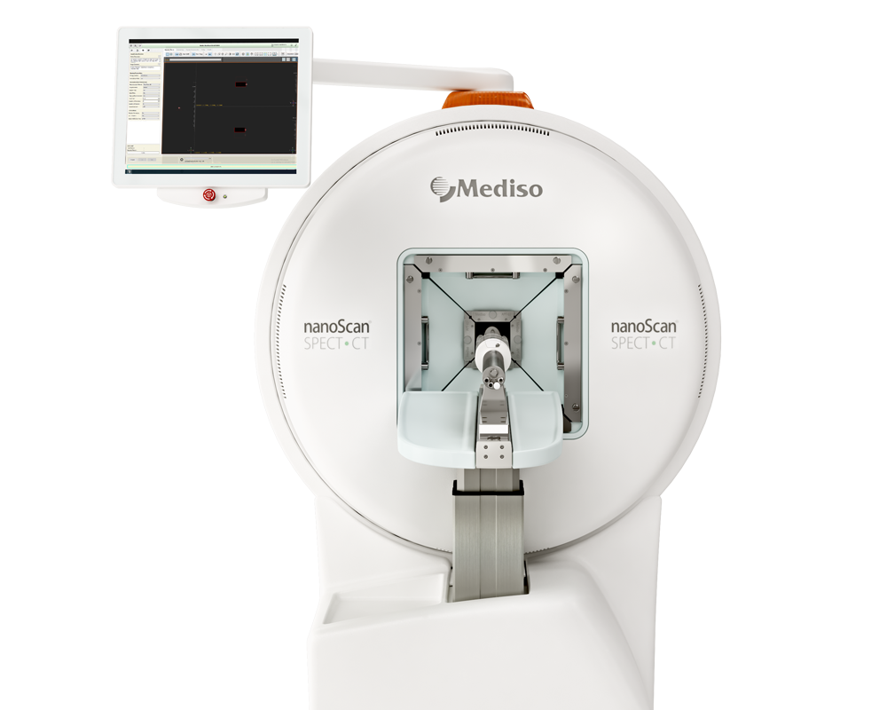
Results from the nanoScan SPECT/CT and nanoScan PET/CT
PET/CT images were acquired with a nanoScan® PET/CT (Mediso Medical Imaging Systems, Budapest, Hungary) scanner operating in list mode using a 400–600 keV energy window and a coincidence window of 1:3. CT scans were acquired for anatomical reference and attenuation correction (55 keV X-ray, exposure time 1000 ms, and 360 projections and pitch 1). PET projection data were reconstructed using the Tera-tomo® software package provided with the scanner —a Monte Carlo-based fully 3D iterative algorithm with four iterations, six subsets, and 0.4 mm isotropic voxel size; corrections for attenuation, scatter, and dead-time were enabled. The data were then visualised and quantified using VivoQuant© v.2.50 (InviCro, Boston, MA, USA) software. SPECT/CT imaging was performed on a NanoSPECT/CT Silver Upgrade scanner (Mediso; 4 heads, 4 × 9 1.0 mm multipinhole collimators) in helical scanning mode using energy windows centred around 93.20 ± 20% keV (primary), 184.60 ± 20% keV (secondary), and 300.00 ± 20% keV (tertiary). CT images were acquired using 45 kVp tube voltage and 1000 ms exposure time in 180° projections. SPECT/CT data sets were reconstructed using the HiSPECT 1.4.2611 (SciVis, Gottingen, Germany) reconstruction software package using standard reconstruction with 35% smoothing and 9 iterations. Images were coregistered and analysed using VivoQuant v2.50 (InviCro, Needham, MA, USA).
Figure 4. shows the biodistribution of [68Ga]KP46 and [68Ga]Ga-acetate, each at tracer (no-carrier-added) concentrations, was determined in NSG mice bearing A375 human melanoma xenografts over a period of four hours (a limit imposed by the 68 min half-life of 68Ga) after intravenous administration, by PET/CT imaging. Injection of [68Ga]gallium acetate led to the visible uptake of 68Ga in the tumour by four hours, rising to ca. 6% ID/g. This was accompanied by increasing uptake in the joints and urinary bladder during this period (Figure 4A)—a biodistribution pattern typical of 67/68Ga salts. Injection of [68Ga]KP46, on the other hand, led to no visible tumour accumulation of 68Ga during the four-hour PET/CT scan post-i.v. injection; instead, radioactivity was taken up largely in the liver, with subsequent translocation into the intestines, and in the myocardium (Figure 4B).
Figure 5. PET/CT images showed an uptake pattern consistent with rapid transit from stomach to small and large intestine. Despite the expectation that the liver would be the main initial repository (delivered via the portal circulation) for radioactivity absorbed in the intestines, very little liver activity, or indeed activity in any other tissues, was observed.
Because the time scale of in vivo experiments with 68Ga was limited by its short half-life (68 min), investigations of the biodistribution of orally administered KP46 were repeated using the longer-lived (78.3 h) 67Ga gamma-emitting radionuclide 67Ga. SPECT imaging with 67Ga enabled the in vivo fate of gallium to be tracked for 4, 24, and 48 h, both at tracer-only level (group E) and bulk pharmacological level (group F). In both groups, SPECT/CT images at the four-hour time point were consistent with the four-hour [68Ga]KP46 PET study described above, with radioactivity largely confined to the stomach and intestines. At 24 h, however, significant differences emerged between the “tracer level” group and the “bulk level” group. In the “tracer level” group (E), SPECT/CT imaging showed little radioactivity in any organs, and the vast majority of radioactivity had been excreted in faeces (as evident from both the SPECT scans and radioactivity measurements of the cage contents as well as animal tissues). In the “bulk level” group (F), on the other hand, faecal excretion was significantly delayed, and a much higher fraction of radioactivity remained in the body. This delay allowed time for absorption of some 67Ga from the gut and translocation to other tissues; although substantial radioactivity remained in the intestines, measurable amounts appeared in the liver, joints, and tumour (Figure 7).
Mice in the “bulk level” group were imaged again 48 h postadministration of the [67Ga]KP46. By this time, although much of the radioactivity had been excreted in faeces, the images showed visible uptake in tumour, liver, and joints (Figure 8.).
- In summary, the authors could conclude that the use of radioactive gallium (68Ga and 67Ga), particularly in conjunction with PET and SPECT imaging, respectively, has a significant contribution to make in elucidating the absorption and trafficking of gallium drugs. Indeed, subject to dosimetry and regulatory evaluation, there are no significant barriers to extending imaging experiments from mice to humans as part of future clinical trials, since clinically usable 68Ga and 67Ga and clinical PET and SPECT scanners are widely available.
- Using 68Ga and 67Ga as tracers, the authors showed that when KP46 enters the circulation intact, the liver and myocardium are the primary targets, whereas ionic gallium primarily targets the joints and tumour. The biodistribution of gallium following oral administration of KP46 shows extremely inefficient absorption of gallium, and the biodistribution of the small fraction that is absorbed matches that of i.v.-administered ionic gallium and not that of i.v.-administered KP46.
- Hence, it can be concluded that the gallium arriving at the tumour is not in the form of KP46. In vitro protein-binding studies with radiogallium combined with octanol extraction studies of the stomach and intestine contents suggest that the dissociation occurs not in the blood but in the gut.
Comment pouvons-nous vous aider?
N'hésitez pas à nous contacter pour obtenir des informations techniques ou à propos de nos produits et services.
Contactez-nous

