Loss of TREM2 rescues hyperactivation of microglia, but not lysosomal deficits and neurotoxicity in models of progranulin deficiency
2022.01.12.
Anika Reifschneider et al., The EMBO Journal, 2022
Summary
Neurodegenerative diseases are currently incurable and novel therapeutic strategies are desperately required. Besides disease-defining protein deposits, microgliosis is observed in almost all neurodegenerative diseases. Microgliosis can be detrimental. However, recent findings strongly suggested that certain microglial responses to brain pathology may also be neuroprotective. This is based on the identification of variants in genes predominantly or exclusively expressed in microglia within the brain that increase the risk for late-onset Alzheimer’s disease (LOAD) and other neurodegenerative disorders. Protective microglial functions became particularly evident upon functional investigations of coding variants found within the triggering receptor expressed on myeloid cells 2 (TREM2) gene, which can increase the risk for LOAD and other neurogenerative disorders including frontotemporal dementia-like syndromes. These TREM2 variants reduce lipid ligand binding, lipid and energy metabolism, chemotaxis, survival/proliferation, phagocytosis of cellular debris, and potentially other essential microglial functions. Moreover, a loss of TREM2 function locks microglia in a dysfunctional homeostatic state, in which they are unable to respond to pathological challenges by inducing a disease-associated mRNA signature.
Disease-associated microglia (DAM) respond to amyloid pathology by clustering around amyloid plaques where they exhibit a protective function by encapsulating the protein deposits via a barrier function that promotes amyloid plaque compaction and reduces de novo seeding of amyloid plaques. TREM2 is therefore believed to be a central target for therapeutic modulation of microglial functions. A number of agonistic anti-TREM2 antibodies were recently developed, which either enhance cell surface levels of signaling-competent TREM2 by blocking TREM2 shedding and/or crosslinking TREM2 receptors to stimulate downstream signaling via Syk phosphorylation. In preclinical studies, these antibodies boost protective functions of microglia as shown by enhanced amyloid β-peptide and myelin clearance, reduced amyloid plaque load, improved memory in models of amyloidosis, and supported axon regeneration and remyelination in models of demyelinating disorders such as multiple sclerosis.
Although increased TREM2 may be protective in AD patients, in other neurodegenerative diseases, microglia may be overactivated and become dysfunctional. Therefore, in these contexts, antagonistic TREM2 antibodies may display therapeutic benefit through dampening microglial hyperactivation. A well-described example of a neurodegenerative disorder where microglia are hyperactivated is GRN-associated frontotemporal lobar degeneration (GRN-FTLD) with TDP-43 (transactive response DNA-binding protein 43 kDa) deposition caused by progranulin (PGRN) deficiency. In models of GRN-FTLD-associated haploinsufficiency, hyperactivation of microglia is evident, as demonstrated by an increased disease-associated mRNA signature as well as strongly increased 18-kDa translocator protein positron emission-tomography ((TSPO)-PET) signals in mouse models. This is the opposite phenotype of Trem2 knockout (KO) microglia, which are locked in a homeostatic state. Hyperactivation of microglia is also observed in the brain of GRN-FTLD patients. PGRN is a secreted protein, which is also transported to lysosomes, where it appears to control activity of hydrolases, such as cathepsins and glucocerebrosidase (GCase). Total loss of PGRN results in a lysosomal storage disorder. A potential synergistic contribution of lysosomal dysfunction and hyperactivated microglia to the disease pathology and specifically to the deposition of TDP-43 in neurons is likely but currently not understood.
To determine whether hyperactivation of microglia and its pathological consequences in Grn KO mice are dependent on aberrant TREM2 signaling, the authors sought to reduce the microglial activation status by crossing them to Trem2 KO mice. This reduced the expression of DAM genes, suggesting that negative modulation of TREM2 signaling may be exploited to lower microglial activation in neuroinflammatory disorders. In analogy to the agonistic 4D9 TREM2 antibody developed earlier in the authors' laboratory, they therefore generated monoclonal antibodies with opposite, namely antagonistic, properties.
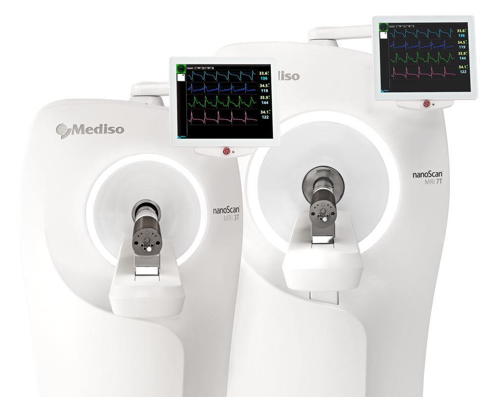
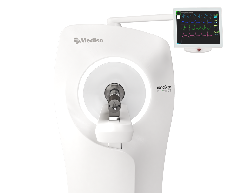
Results from the nanoScan PET/MRI 3T
All mice were scanned with the nanoScan PET/MRI 3T scanner with a single-mouse imaging chamber. A 15-min anatomical T1 MR scan was performed at 15 min after [18F]-FDG injection or at 45 min after [18F]-GE180 injection (head receive coil, matrix size 96 × 96 × 22, voxel size 0.24 × 0.24 × 0.80 mm³, repetition time 677 ms, echo time 28.56 ms, and flip angle 90°). PET emission was recorded at 30–60 min p.i. ([18F]-FDG) or at 60–90 min p.i. ([18F]-GE-180). PET list-mode data within 400–600 keV energy window were reconstructed using a 3D iterative algorithm (Tera-Tomo 3D, Mediso Ltd) with the following parameters: matrix size 55 × 62 × 187 mm³, voxel size 0.3 × 0.3 × 0.3 mm³, eight iterations, six subsets. Decay, random, and attenuation correction were applied. The T1 image was used to create a body–air material map for the attenuation correction. The authors studied PET images of Grn KO mice (n = 8), Trem2 KO mice (n = 10 or n = 9), Double KO mice (n = 10), and WT mice (n = 15), all female at an average age of 10.9 ± 1.6 months or 11.1 ± 1.6 months, as indicated in the figure legends. Normalization of injected activity was performed by the previously validated myocardium correction method for [18F]-GE-180 TSPO-PET and by standardized uptake value (SUV) normalization for [18F]-FDG-PET. Groups of Grn KO, Trem2 KO, and Double KO mice were compared against WT mice by calculation of %-differences in each cerebral voxel. Finally, [18F]-TSPO-PET and [18F]-FDG-PET values deriving from a whole-brain VOI were extracted and compared between groups of different genotypes by a one-way ANOVA including Tukey post hoc correction.
Figure 1. shows TSPO PET imaging, which indicates rescue of microglial hyperactivation in Double−/− mice. Fig. 1A.: Axial slices as indicated below (A1, A2, and A3) show %-TSPO-PET differences between Grn−/−, Trem2−/−, or Double−/− mice and WT at the group level. Images adjusted to an MRI template indicate increased microglial activity in the brain of Grn−/− mice (hot color scale), compensated microglial activity in the brain of Double−/− mice, and decreased microglial activity in the brain of Trem2−/− mice (cold color scale), each in contrast against age-matched WT mice. Fig. 1B.: Scatter plot illustrates individual mouse TSPO-PET values derived from a whole-brain volume of interest. A total of 8–15 female mice per group at an average age of 11.1 ± 1.6 months (Grn−/− (n = 8), Trem2−/− (n = 9), Double−/− (n = 10), and WT (n = 15)). Data represent mean ± SD. For statistical analysis, one-way ANOVA with Tukey post hoc test was used. Statistical significance was set at *P < 0.05; **P < 0.01; ns, not significant.
Figure 9. Hyperactivation of microglia in Grn−/− mice is not deleterious. Fig. 9E.: The same cohort of mice scanned for TSPO-PET was additionally scanned for FDG-PET. Axial slices as indicated show %-FDG-PET differences among Grn−/−, Trem2−/−, and Double−/− (all cold color scales) when compared to WT at the group level. Images were adjusted to an MRI template. Fig. 9F.: Bar graph illustrates individual FDG-PET values derived from a whole-brain volume of interest. Data represent mean ± SD. A total of 8–15 female mice per group at an average age of 10.7 ± 1.5 months (Grn−/− n = 8, Trem2−/− n = 10, Double−/− n = 10, WT n = 15) were used.
- Eliminating TREM2 function and thus reducing hyperactivation by two independent approaches do not rescue lysosomal dysfunction caused by GRN deficiency, but rather exacerbates pathological endpoints characteristic for neurodegeneration, including elevation of CSF NfL and reduced transcription of the neuroprotective transcription factor Npas.
- These findings suggest that TREM2-dependent microglia hyperactivation in models of GRN deficiency does not promote neurotoxicity, but rather neuroprotection.
How can we help you?
Don't hesitate to contact us for technical information or to find out more about our products and services.
Get in touch
