Preclinical evaluation of FAP-2286 for fibroblast activation protein targeted radionuclide imaging and therapy
2022.05.24.
Dirk Zboralski et al., European Journal of Nuclear Medicine and Molecular Imaging, 2022
Summary
Fibroblast activation protein (FAP) is a single-pass type II transmembrane glycoprotein, with a large extracellular domain composed of an α/β-hydrolase and an 8-bladed β-propeller. As a member of the S9 prolyl oligopeptidase family, FAP has post-proline exopeptidase activity similar to dipeptidyl peptidase 4 (DPP4), and an additional endopeptidase activity like prolyl endopeptidase (PREP).
FAP is expressed during embryonic development, with highly restricted expression in healthy adult tissues. However, FAP upregulation can occur in diseases associated with activated stroma including wound healing, rheumatoid arthritis, cirrhosis, pulmonary fibrosis, and solid cancers. In most epithelial cancers, FAP is selectively expressed on the cell surface of cancer-associated fibroblasts (CAFs) present in the tumor microenvironment. These CAFs play important roles in tumor growth and metastasis through their effects on angiogenesis, the extracellular matrix, and the immune system. In addition, in several tumors of mesenchymal origin, notably sarcoma and mesothelioma, FAP expression has also been observed on the neoplastic cells themselves and is similarly associated with promoting tumor progression and metastasis. Given its restricted expression profile and functions, FAP is a promising target for the selective delivery of anticancer therapies to tumors of a broad range of cancer indications.
Many FAP-targeting agents have been developed including small molecule inhibitors and immunoglobulins. However, antibodies are well known to have an extended blood circulating half-life which may elicit greater normal tissue toxicity, particularly in the bone marrow when conjugated to a radionuclide. In contrast, small molecular weight radioconjugates have the advantage of rapid tumor delivery coupled to fast systemic clearance to potentially circumvent this toxicity.
One approach to targeting FAP is through radiolabeled theranostic agents, integrating both noninvasive disease diagnosis and treatment. Validated targeted radiopharmaceuticals include LUTATHERA® (177Lu-dotatate; Advanced Accelerator Applications), a cyclic peptide targeting somatostatin subtype 2 receptor (SSTR2) armed with the β-emitting radionuclide lutetium-177 (177Lu), which has been approved for treatment of SSTR2-positive patients with gastroenteropancreatic neuroendocrine tumors (GEP-NET); and 177Lu-PSMA-617, a small molecule targeting prostate-specific membrane antigen (PSMA) linked to 177Lu, which recently met both primary survival endpoints in the phase III VISION trial for PSMA-positive metastatic castration-resistant prostate cancer (mCRPC).
FAP is an emergent target in the radiopharmaceutical field and has been referred to as “the next billion dollar nuclear theranostics target”. This description originates from the development of the FAP inhibitor (FAPI) series of small molecule quinoline-based radiotracers that have shown high specificity across various primary and metastatic tumors. The diagnostic utility of 68Ga-FAPI–based positron emission tomography (PET) has been established in various cancer types, demonstrating substantial accumulation in cancer of unknown primary (CUP), sarcoma, esophageal cancer, breast cancer, and cholangiocarcinoma. In addition, 68Ga-FAPI-04 appears to outperform 18F-fluorodeoxyglucose (18F-FDG) in discriminating the primary and distant metastatic lesions in certain indications.
Further structural modifications of the FAPI series of compounds are currently ongoing to improve pharmacology and allow the attachment of alternative radionuclides. In parallel, multiple academic and commercial entities have recently described highly related FAP-targeting molecules with similar results, including the lead therapeutic compound FAPI-46 with a methylamino alteration in the linker region. However, the FAPI radiotracers are established around UAMC1110, a small molecule inhibitor of FAP, and exhibit fast clearance from tumors, limiting their potential therapeutic effectiveness.
To address the low tumor retention of the FAPI series, the authors developed the FAP-2286 compound, based on a novel class of FAP-targeting modalities, that utilizes cyclic peptides as binding motifs. Cyclic peptides are known to have potentially improved biological properties over linear counterparts, including greater binding affinity and selectivity due to their conformational rigidity and increased plasma stability. FAP-2286 is composed of such a cyclic peptide, which is linked to a tetraazacyclododecane tetraacetic acid (DOTA) allowing chelation of radionuclides for imaging or therapeutic applications. For imaging, the radionuclides gallium-68 (68Ga) or indium-111 (111In) were chelated to FAP-2286 for use as a PET or single-photon emission computed tomography (SPECT) imaging agent, respectively. For therapeutic use, 177Lu was chelated to FAP-2286. Herein, the in vitro and in vivo characterization of FAP-2286 and its metal complexes are reported and compared to FAPI-46.
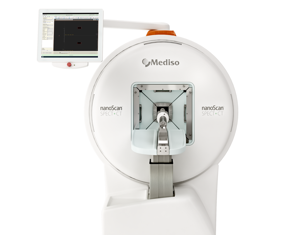
Results from the nanoScan SPECT/CT and nanoScan PET/CT
For the imaging studies, thirty megabecquerels (30 MBq/nmol) 111In-FAP-2286 (n = 9) or 111In-FAPI-04 (n = 6) was injected intravenously (IV) into HEK-FAP tumor-bearing mice and the distribution of the radiotracers was assessed using the NanoSPECT/computed tomography (CT) system. For PET imaging, 10 MBq of 68Ga-FAP-2286 or 68Ga-FAPI-46 (10 MBq/nmol) was given IV into HEK-FAP tumor-bearing mice (n = 3) followed by PET/CT analysis using the nanoScan PET/CT system. For SPECT imaging of 177Lu-labeled FAP-2286 or 177Lu-labeled FAPI-46, 30 MBq was administered IV (30 MBq/nmol; n = 3) and the distribution of the radiotracers was assessed using the nanoScan SPECT/CT system.
Fig. 4 shows the biodistribution of 111In-FAP-2286 was evaluated in vivo by SPECT imaging following a single dose of 30 MBq in HEK-FAP tumor bearing mice. The radiotracer was rapidly enriched within the HEK-FAP xenografts with low off-target accumulation and predominantly renal elimination (Fig. 4A). High tumor-to-background signal ratio was observed from 1 h post injection (p.i.) onward. At 1 h p.i. of 111In-FAP-2286, tumor uptake was 11.1 percent injected dose per gram (%ID/g). Accumulation of 111In-FAP-2286 was stably maintained in the tumors with 9.1%ID/g at 48 h p.i. (Fig. 4B, Supplementary Table S2). Normal tissues including liver, salivary gland, and shoulder showed very limited uptake. The organ with the highest non-target uptake was the kidney; however, an increasing tumor-to-kidney (T/K) ratio was observed over time with the highest differential uptake of 7.5 T/K obtained at 48 h p.i. (Supplementary Table S2).
Fig 7. shows the comparison of the biodistribution of FAP-2286 against FAPI-46, the current lead therapeutic compound from the FAPI radiotracer series. 68Ga-FAP-2286 and 68Ga-FAPI-46 were rapidly enriched within the HEK-FAP xenografts with predominantly renal elimination (Fig. 7A, Supplementary Table S3). High tumor specificity was observed at all 3 timepoints assessed (0.5, 1, and 3 h p.i.). At 0.5 h after administration of 68Ga-FAP-2286 and 68Ga-FAPI-46, tumor uptake was 9.8 and 9.3 %ID/g, respectively. Accumulation of 68Ga-FAP-2286 and 68Ga-FAPI-46 was maintained at 3 h after injection with 10.8 and 9.2 %ID/g, respectively, and no significant difference in tumor distribution was observed between both compounds (Fig. 7C).
The 177Lu-radiolabeled FAP-2286 and FAPI-46 also demonstrated rapid accumulation within the HEK-FAP xenografts with low background signal at 3 h after dosing (Fig. 7B, Supplementary Table S3). However, significantly higher tumor retention of 177Lu-FAP-2286 compared to 177Lu-FAPI-46 was observed at 24 and 72 h p.i. (P < 0.001). Tumor uptake of 177Lu-FAP-2286 at the 3-h timepoint was 21.1 %ID/g and demonstrated durable retention with 16.4 %ID/g 72 h later. The time-integrated activity coefficient (TIAC) and absorbed dose were calculated to be 11.2 MBq*h/MBq and 2.8 Gy/MBq, respectively. In contrast, 177Lu-FAPI-46 accumulation decreased from 15.3 %ID/g at the 3-h timepoint to 1.6 %ID/g after 72 h resulting in a lower TIAC of 0.9 MBq*h/MBq and absorbed dose of 0.3 Gy/MBq (P < 0.001, Fig. 7D, Supplementary Table S3 and Fig. S4). Similar to the gallium-labeled compounds, normal tissues showed limited uptake and rapid clearance of the radiotracers with the highest level in the kidneys. A higher kidney accumulation was observed for 177Lu-FAP-2286 compared to 177Lu-FAPI-46 with the greatest difference at 3 h after injection with 2.2 %ID/g and 0.7 %ID/g, respectively, which decreased to 0.6 %ID/g and 0.2 %ID/g, respectively, at 72 h p.i. (Supplementary Table S3).
- Taken altogether, FAP-2286 demonstrates compelling characteristics of a targeting agent with potent and selective FAP binding that leads to high tumor accumulation and substantial therapeutic efficacy.
- The preclinical studies as well as the first-in-human experience support further development of 68Ga-FAP-2286 and 177Lu-FAP-2286, and evaluation in patients with advanced solid cancers in the ongoing phase 1/2 clinical study LuMIERE (NCT04939610) for assessment of their safety, pharmacokinetics, dosimetry, and efficacy.
W czym możemy pomóc?
Skontaktuj się z nami aby uzyskać informacje techniczne i / lub wsparcie dotyczące naszych produktów i usług.
Napisz do nas

