Molecular Imaging and Preclinical Studies of Radiolabeled Long-Term RGD Peptides in U-87 MG Tumor-Bearing Mice
2021.05.21.
Wei-Lin Lo et al., International Journal of Molecular Sciences, 2021
Summary
Cancer is the second leading cause of death in the world. In recent years, integrin-mediated biological activity targeted against cancer has been improving. Mammals have 24 different integrins, consisting of 18 α-subunits and 8 β-subunits, which are cell adhesion receptors connected to the extracellular matrix (ECM) and other cells. Integrins participate in signal transduction pathways to regulate cell growth, migration, motility, proliferation and survival. The αvβ3 integrin is overexpressed in new tumor blood vessels and various tumor cells including neuroblastoma, osteosarcoma, melanoma, glioblastoma, breast cancer and lung cancer. Furthermore, integrin plays important roles in cancer proliferation, invasion, survival, metastasis and angiogenesis. According to the above characteristics, integrin is developed as a suitable target for cancer diagnosis and therapy.
The Arg–Gly–Asp (RGD) peptide is expressed on ECM and recognized by integrin, and shows a high affinity for αvβ3 integrin. The RGD-containing peptides can be divided into linear and cyclic forms in structure, and have different modifications for different purposes, such as to increase binding affinity, extend half-life, combine with another drug, etc. Cyclization of RGD can enhance biological activity and significantly improve its selectivity and binding ability to receptors, and the cyclic conformational structure can also prevent proteolysis. The cyclic Arg–Gly–Asp–D–Phe–Lys (cRGDfK) sequence can inhibit the adhesion of fibronectin to cells, and the binding affinity to integrin αvβ3 is much higher than the linear RGD peptide sequence.
In all molecular imaging modalities, positron emission tomography (PET) and single photon emission computed tomography (SPECT) occupy a specific location, using high-affinity and high-specific molecular radiotracers as imaging probes to visualize and measure physiological processes. Targeting peptides are combined with different radionuclides, diagnostic or therapeutic isotopes, and are often used as radiotracers. The radiolabeled RGD peptide has been studied for cancer imaging and radionuclide therapy. Recently, Chen et al. published a novel EB-RGD for imaging and radiotherapy, consisting of a truncated Evans blue dye (EB) molecule with the binding capacity of albumin, a metal chelate that allows radiolabeling, and RGD peptide that binds to integrin. The micromolar affinity and reversible binding of EB derivatives to albumin extend the half-life of the drug in the blood. The EB-conjugated drug can improve pharmacokinetic properties and prolongs blood circulation. Moreover, EB-RGD may become an effective treatment option for targeted radionuclide therapy (TRT) by labeling with the β-emitting radionuclide lutetium-177 (177Lu).
In this research, the authors developed a novel long-circulation integrin-targeted molecule DOTA-EB-cRGDfK based on EB-RGD. To increase tumor accumulation and retention for radioligand therapy, and reduce dosage of radionuclide, the authors designed and conjugated an EB molecule and DOTA chelator onto RGD peptide and labeled it with indium-111(111In). These studies selected U-87 MG human glioblastoma with a high expression of αvβ3 integrin as the model.
Results from nanoScan SPECT/CT
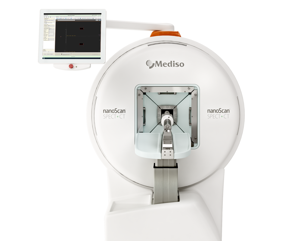
For the imaging studies, each mouse was tail-vein injected with about 18.5 MBq and 2.5 μg (50–100 μL) of radiolabeled 111In-DOTA-EB-cRGDfK, 111In-DOTA-cRGDfK, or 111In-DOTA-EB. For the blocking study, animals were pre-treated with a 120-fold molar excess (300 μg) DOTA-EB-cRGDfK by tail vein injection 10 min ago. SPECT images and X-ray CT images were acquired using a nanoSPECT/CT scanner system. The mice were anesthetized with 1–2% isoflurane during the imaging acquisition. The imaging acquisition was accomplished at 60 s per frame. The energy window was set at 171 and 245 KeV ± 10%, the image size was set at 256 × 256, and the field of view of 60 mm × 100 mm. For image reconstruction, the HiSPECT and Nucline software were used for the SPECT and CT images, respectively. The InVivoScope software was used for the fusion of SPECT and CT images. The image quantification of the region of interest (ROI) was acquired by PMOD v. 3.3 (Zürich, Switzerland). The SPECT images were presented on a scale of 2.5% ID/g to 25% ID/g.
Figure 3. presents nanoSPECT/CT images of U-87 MG xenografts in mice administered with different radiolabeled peptides. After injection of 111In-DOTA-EB-cRGDfK, tumor accumulation can be found within 0.5 h, and gradually increased with time and there was still a clear signal until 72 h. 111In-DOTA-EB-cRGDfK showed significantly higher tumor uptake than control peptides at all time points by image quantification results (12.36 ± 0.88, 16.59 ± 2.16, 18.66 ± 2.15, 25.09 ± 4.76, 25.49 ± 3.88 and 23.61 ± 2.98% ID/g in tumor at 0.5, 2, 4, 24, 48 and 72 h after injection, respectively). When blocking with 120-fold molar excess of DOTA-EB-cRGDfK, the image showed significantly inhibitive binding of 111In-DOTA-EB-cRGDfK in tumors (5.41 ± 1.24, 5.69 ± 1.31, 5.84 ± 0.88, 7.13 ± 2.62, 8.19 ± 2.22 and 7.52 ± 2.35% ID/g in tumors at 0.5, 2, 4, 24, 48 and 72 h after injection, respectively). Control peptide 111In-DOTA-cRGDfK showed low tumor uptake and rapid excretion from the body. Comparing with 111In-DOTA-EB-cRGDfK, the highest tumor uptake of 111In-DOTA-cRGDfK was 2.0 ± 0.5% ID/g at 0.5 h. In addition, 111In-DOTA-EB showed relatively obvious radiation signals in non-tumor organs such as muscle and liver. The ratios of tumor to muscle and tumor to liver were 1.8 ± 0.7 and 0.5 ± 0.1 at 0.5 h, respectively. The highest tumor uptake of 111In-DOTA-EB was 10.3 ± 4.4% ID/g at 24 h, and it slightly dropped to 9.97 ± 4.37% ID/g at 72 h.

- The biodistribution results showed significant tumor accumulation (27.1 ± 2.7% ID/g) and the tumor to non-tumor ratio was 22.85 at 24 h after injection.
- 111In-DOTA-EB-cRGDfK shows favorable pharmacokinetics. The radiolabeled peptides usually showed fast blood clearance, but the T1/2λz of 111In-DOTA-EB-cRGDfK was 77.3 h. The extended half-life may increase the fully useful dosage.
- DOTA is a good chelator for labeling different radionuclides, such as 111In for SPECT imaging and 177Lu for treatment.
Comment pouvons-nous vous aider?
N'hésitez pas à nous contacter pour obtenir des informations techniques ou à propos de nos produits et services.
Contactez-nous
