Patient-derived xenografts and in vitro model show rationale for imatinib mesylate repurposing in HEY1-NCoA2-driven mesenchymal chondrosarcoma
2021.11.26.
Polona Safaric Tepes, Danilo Segovia, Sania Jevtic, Daniel Ramirez, Scott K. Lyons and Raffaella Sordella
nature>laboratory investigation, 2021 Nov
Abstract:
Mesenchymal chondrosarcoma (MCS) is a high-grade malignancy that represents 2–9% of chondrosarcomas and mostly affects children and young adults. HEY1-NCoA2 gene fusion is considered to be a driver of tumorigenesis and it has been identified in 80% of MCS tumors. The shortage of MCS samples and biological models creates a challenge for the development of effective therapeutic strategies to improve the low survival rate of MCS patients. Previous molecular studies using immunohistochemical staining of patient samples suggest that activation of PDGFR signaling could be involved in MCS tumorigenesis. This work presents the development of two independent in vitro and in vivo models of HEY1-NCoA2-driven MCS and their application in a drug repurposing strategy. The in vitro model was characterized by RNA sequencing at the single-cell level and successfully recapitulated relevant MCS features. Imatinib, as well as specific inhibitors of ABL and PDGFR, demonstrated a highly selective cytotoxic effect targeting the HEY1-NCoA2 fusion-driven cellular model. In addition, patient-derived xenograft (PDX) models of MCS harboring the HEY1-NCoA2 fusion were developed from a primary tumor and its distant metastasis. In concordance with in vitro observations, imatinib was able to significantly reduce tumor growth in MCS-PDX models. The conclusions of this study serve as preclinical results to revisit the clinical efficacy of imatinib in the treatment of HEY1-NCoA2-driven MCS.
Results from the nanoScan® PET/CT
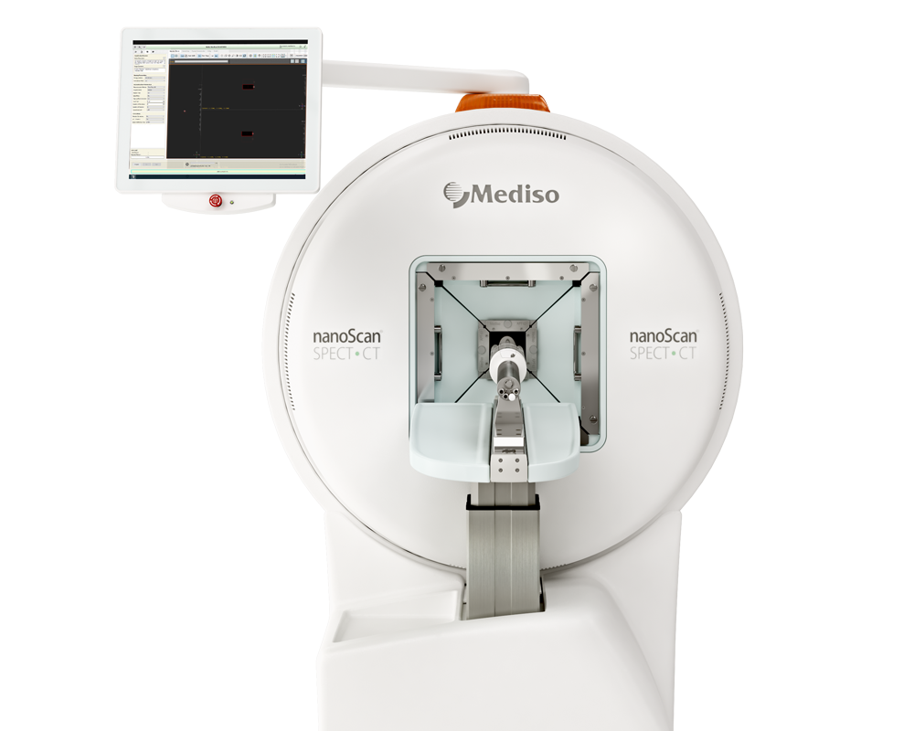
MicroCT scan were aquired by nanoScan PET/CT (Mediso) at an energy of 50 kVp and exposure of 186 μAs, with 480 projections per bed position. Tomographic images were reconstructed to an isotropic voxel size of 250 μm using a filtered back-projection algorithm with a high-resolution Ram-Lak filter. Interview Fusion software (Mediso) was used for CT image post processing and VivoQuant (Invicro) was used to manually segment tumors and measure tumor volume and mean density.

a, A representative reconstructed 3D image of CT imaging of PDX-primary tumors treated with vehicle. The magenta color represents the cartilaginous component of the primary tumor b, A representative reconstructed 3D image of CT imaging of PDX-primary tumors treated with imatinib

f A representative reconstructed 3D image of CT imaging of PDX metastasis treated with vehicle. The magenta color represents the cellular component of the tumor, white color shows the cartilaginous component. g A representative reconstructed 3D image of CT imaging of PDX metastasis treated with imatinib. The magenta color represents the cellular component of the tumor, white color shows the cartilaginous component.
Comment pouvons-nous vous aider?
N'hésitez pas à nous contacter pour obtenir des informations techniques ou à propos de nos produits et services.
Contactez-nous

