Myocardial perfusion recovery induced by an α-calcitonin gene-related peptide analogue
2021.06.04.
Simon Bentsen MD et al., Journal of Nuclear Cardiology, 2021
Summary
Myocardial infarction (MI) is the leading cause of heart failure, despite the introduction of primary percutaneous coronary intervention (PCI). PCI has improved survival after an MI, but the recovering myocardium is still impaired after PCI. Therefore, it is crucial to develop new treatments to protect and improve myocardial recovery after an MI.
After the onset of an MI, there is insufficient oxygen supply to the myocytes. To protect the myocytes prior to PCI, it is crucial to reduce cardiac workload. Vasodilation of the coronary arteries, the peripheral arterial bed, and venous capacitance vessels is a known and used method of reducing afterload and the myocardial workload. However, peripheral vasodilation can increase the workload of the heart because of compensatory elevated heart rate, which could lead to increased ischemic damage. Calcitonin gene-related peptide (CGRP) is one of the most potent vasodilators known.
Exogenous CGRP has been shown to induce cardioprotective effects in isolated hearts and CGRP antagonism reduces the cardioprotective effects. Also, exogenous CGRP induces positive chronotropic and inotropic effects on isolated guinea pig hearts and isolated human myocardial trabeculae. In three clinical studies with CGRP infusion for the treatment of cerebral vasospasm, adverse effects of increased heart rate and peripheral hypotension were observed, and the studies were stopped before completion.
Recently, a metabolically stable CGRP analogue, SAX, with a lipophilic tail has been synthesized. This analogue reverses hypertension and complications of hypertension in experimental animal studies. Data show that SAX and CGRP display similar pharmacological actions, but the potency of peripheral vasodilation induced by SAX is approximately 10 times lower than native CGRP.
The aim of this study was to examine a potential cardioprotective effect of SAX in rats undergoing experimental acute myocardial infarction by permanent left anterior descending (LAD) occlusion. The authors hypothesized that SAX will increase perfusion of the acutely hypoperfused area without promoting increased workload on the heart and that this will cause improved recovery of the myocardial perfusion.
Results from the nanoScan SPECT/CT
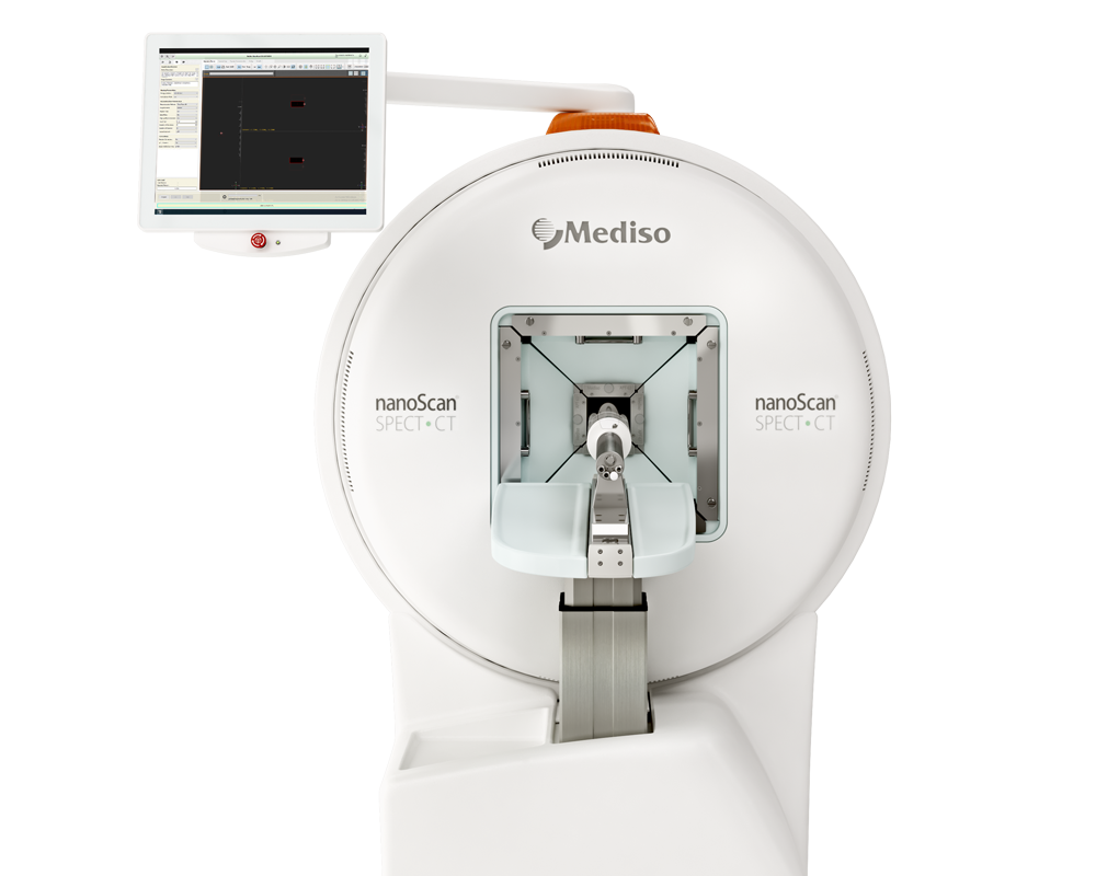
The rats underwent acute SPECT/CT scan one hour after surgery (to determine baseline non-perfused area). In Sprague-Dawley rats, [99mTc]Tc-sestamibi clears from the blood rapidly and is taken up in the heart with very little redistribution. Therefore, the injection of [99mTc]Tc-sestamibi 20 min before injection of SAX or placebo enables the detection of the non-perfused area before any potential impact of the first SAX injection. Three weeks after surgery, the rats were follow-up scanned with SPECT/CT. This enabled a determination of final infarct size. All imaging was performed on the Mediso nanoScan SPECT/CT scanner.
The rats were anesthetized with sevoflurane 4% for the SPECT/CT scan. On the day of the surgery, the rats were perioperatively injected with [99mTc]Tc-sestamibi (median 106 mBq[99; 116]), as described above. Twenty-one days after surgery, the rats were anesthetized with sevoflurane 4%. A 24G intravenous catheter was placed in the tail vein (Vasofix Safety, Braun, Denmark). The rats were then injected with [99mTc]Tc-sestamibi (median 122 mBq[106; 129]). During the SPECT/CT scan, the rats were placed on MultiCell Rat bed, in a prone position. The rats were monitored by way of electrocardiography (ECG), respiratory rate, and core temperature. A computed tomography (CT) scout image was obtained to ensure correct positioning of the SPECT detector’s field of view over the heart. One hour after injection of [99mTc]Tc-sestamibi, the SPECT acquisition was initiated. Scan time was between 20 and 40 min, depending on injected activity, adjusted to ensure a number of counts above 100,000 counts/frame/detector. For attenuation correction and anatomical co-registration, a CT scan was acquired after the SPECT. All images were reconstructed using vendor software (Mediso Nanoscan, Hungary) and vendor-recommended parameters.
Comment pouvons-nous vous aider?
N'hésitez pas à nous contacter pour obtenir des informations techniques ou à propos de nos produits et services.
Contactez-nous
