Molecular imaging of HER2 expression in breast cancer patients using a novel peptide-based tracer 99mTc-HP-Ark2: a pilot study
2023.01.11.
Jiyun Shi et al., 2023, Journal of Translational Medicine
Summary
Human epidermal growth factor receptor 2-positive (HER2+) breast cancer accounts for 20–30% of invasive breast cancers, and HER2 expression is associated with poor prognosis. HER2+ patients can benefit from HER2-targeted treatment. Trastuzumab (Herceptin®, Genentech, Inc.), as an anti-HER2 monoclonal antibody, is currently the first-line drug for HER2+ breast cancer, as it significantly improves the chances of cure of early HER2+ breast cancer and reduces the risks of recurrence and death. However, the approximately 30% response rate and varying degrees of drug resistance make the accurate diagnosis of HER2 expression essential. In addition, the downregulation of HER2 expression is reported as an indicator of trastuzumab’s antitumor effect. Therefore, HER2 expression assessment is important not only before but also during treatment. In current clinical practice, biopsy-based immunohistochemistry (IHC) and fluorescence in situ hybridization (FISH) are recognized as the gold standards for detecting HER2 status. However, metastases are often found in breast cancer patients, and it is difficult to test the HER2 status of bone metastases by biopsy. It is also difficult for biopsy to identify heterogeneity and discordance of HER2 status between metastases and primary lesions. The temporal and spatial heterogeneity of HER2 expression in breast tumors can lead to inaccurate assessments and further mislead oncologists in choosing the therapeutic regimen. The shortcomings of invasive biopsy analysis have promoted the development of HER2-targeted molecular imaging.
Molecular imaging probes targeting HER2 have been extensively studied. In some clinical studies, the therapeutic antibodies trastuzumab and pertuzumab have been radiolabeled to guide the administration of targeted therapy. However, antibody-based imaging usually needs to wait 2–4 days after administration, which is not convenient to guide treatment decisions in time and expose patients to longer radiation exposure. In addition, radiolabeled antibodies usually have a high liver background uptake, which may lead to poor visualization of liver metastases. Therefore, many small molecule substitutes targeting HER2 have emerged, including antibody fragment, nanobody, affibody and peptide, which have faster blood circulation and better tissue permeability and are more suitable for the development of imaging probes than antibodies, and some of them have been developed as cancer diagnostic probes for clinical trials.
Peptides have many favorable characteristics suitable for the development of imaging agents. In addition to high tissue permeability and fast blood clearance, they are also easy to synthesize and formulate kits, which is more conducive to clinical translation and promotion. However, at present, almost all HER2-targeted peptide probes are in preclinical studies, and clinical translational studies are still scarce. Previously, the authors have developed two probes, 99mTc-H6F and 99mTc-H10F, based on two HER2-targeting peptides that bind to HER2 at different binding sites with trastuzumab (extracellular II vs. extracellular IV) and thus hold the potential to monitor efficacy during trastuzumab therapy. However, 99mTc-H6F has poor water solubility and high lipophilicity, resulting in high gallbladder uptake, which is not conducive to clinical use. 99mTc-H10F has good water solubility, and its ability to monitor treatment efficacy has been verified in animal models. However, its rapid clearance in vivo and relatively low tumor uptake also limit its further clinical application. In this study, the authors developed an improved HER2-targeting molecular probe 99mTc-HP-Ark2 based on H10F peptide through D-shaped amino acids, sequence reversal, dimerization, 8-carbon aliphatic chain and PEG4 chain modification, etc. Compared with the previous probe 99mTc-H10F, 99mTc-HP-Ark2 had enhanced HER2 targeting capability and improved pharmacokinetic properties, thereby increasing its efficacy for the clinical detection of HER2 expression. A pilot prospective clinical study of 99mTc-HP-Ark2 SPECT/CT (single photon emission computed tomography/computed tomography) was performed in breast cancer patients and compared with 18F-FDG PET/CT (positron emission tomography/computed tomography) lesion by lesion.
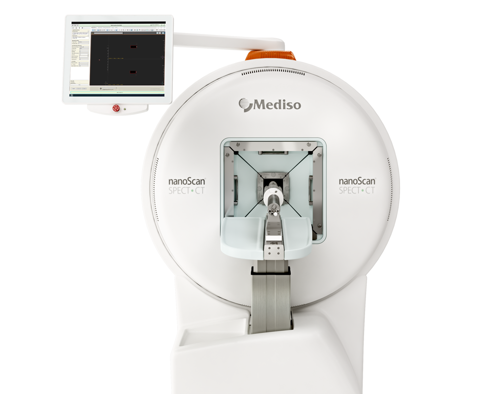
Results from the nanoScan SPECT/CT
The small-animal SPECT/CT imaging of 99mTc-HP-Ark2 was performed in female NOD SCID mice bearing SK-BR-3 human breast cancer xenografts. Each tumor-bearing mouse was injected via the tail vein with ~ 37 MBq (~ 2.5 µg) of 99mTc-HP-Ark2. At 0.5, 1 and 2 h p.i., the mice were anesthetized by inhalation of 2% isoflurane and imaged using a nanoScan SPECT/CT system following a standard protocol. Briefly, the small-animal SPECT images were acquired with the technetium-99 m parameters (peak 140 keV with 20% width, frame time 25 s), and CT images were acquired with default settings (50 kVp, 0.67 mA, and rotation 210°, exposure time 300 ms). All SPECT/CT images were reconstructed and further analyzed by InterView Fusion software, including drawing volumes of interest on tumors and major organs as well as calculating the tumor uptake (%ID/cc) accordingly. Blocking was studied by coinjection of 99mTc-HP-Ark2 with a blocking dose of (25 mg/kg) cold rk peptide or (30 mg/kg) trastuzumab, and nanoScan SPECT/CT imaging was performed at 0.5 h p.i. Small-animal imaging of the monomer tracer 99mTc-HP-rk was also conducted as a control. The in vivo behavior of 99mTc-HP-Ark2 in MCF7, MCF7-HER2, HT29, BxPC3 and MDA-MB-468 tumor xenograft models was also evaluated by nanoScan SPECT/CT imaging at 0.5 h p.i., following the above standard protocol.
Figure 3. shows the in vivo tumor targeting capability of 99mTc-HP-Ark2 was determined in the SK-BR-3 tumor model by nanoScan SPECT/CT imaging. The SK-BR-3 tumors could be clearly visualized at 0.5, 1 and 2 h postinjection (p.i.) (Fig. 3A), even when the tumor was as small as ~ 20 mm3, regardless of whether it was subcutaneous or an in situ model (Additional file 1: Figure S5 & Fig. 3B). Coinjection of excessive cold rk peptide significantly blocked tumor uptake, but excessive cold trastuzumab did not block tumor uptake (Fig. 3B), suggesting that the accumulation of 99mTc-HP-Ark2 in HER2-positive tumors was specifically receptor-mediated and had no cross-interference by trastuzumab. The imaging intensity of 99mTc-HP-Ark2 in tumors was stronger than that of 99mTc-HP-rk (Fig. 3A, B).
The biodistribution results were consistent with the imaging results. 99mTc-HP-Ark2 showed higher tumor uptake than uptake in other organs (except for kidneys) at all three studied time points, 0.5, 1 and 2 h p.i. (Fig. 3C), resulting in a better contrast of tumor-to-background. 99mTc-HP-Ark2 showed notably enhanced tumor uptake (3.99 ± 0.15%ID/g) at 0.5 h p.i. compared to 99mTc-HP-rk (1.94 ± 0.12 %ID/g, P < 0.0001, n = 4), with no significant difference in other organs except the kidneys. In the blocking group, the tumor uptake was significantly blocked by coinjection of excess cold rk peptide (1.42 ± 0.15 %ID/g, P < 0.0001, n = 4), indicating the specific targeting of the probe to HER2-positive tumors (Fig. 3D). In the control groups, the tumor uptake of 99mTc-HP-Ark2 in the HER2-negative MCF7 (1.44 ± 0.08 %ID/g, P < 0.0001, n = 4) and MDA-MB-468 models (1.08 ± 0.06 %ID/g, P < 0.0001, n = 4) was significantly lower than that in the HER2-positive SK-BR-3 model, also indicating the specific targeting of the probe to HER2-positive tumors (Additional file 1: Figure S6). The specificity of Ark2 for HER2 was further verified by immunofluorescence staining of ex vivo tumor tissues. The results showed that Cy5-Ark2 could colocalize with trastuzumab in HER2-positive SK-BR-3 and MCF7-HER2 tumor tissues, while no obvious staining signals were detected in HER2-negative MCF7 tumor tissues (Additional file 1: Figure S7).
- The authors have developed a novel HER2-targeted SPECT imaging probe, 99mTc-HP-Ark2. The kit formulation is simple, efficient, and reproducible, making it very convenient for routine clinical use.
- 99mTc-HP-Ark2 showed enhanced tumor uptake, improved pharmacokinetic properties and increased imaging contrast over the previous 99mTc-H10F probe, which made 99mTc-HP-Ark2 more suitable for clinical application.
- The pilot study in 34 breast cancer patients showed that 99mTc-HP-Ark2 SPECT/CT could noninvasively reflect the status of HER2 in breast cancer, which showed great potential to identify patients to receive trastuzumab treatment and monitor the therapeutic efficacy earlier during trastuzumab treatment.
- This prospective clinical study merits 99mTc-HP-Ark2 SPECT/CT for further clinical validation in larger cohorts.
Comment pouvons-nous vous aider?
N'hésitez pas à nous contacter pour obtenir des informations techniques ou à propos de nos produits et services.
Contactez-nous
