Evaluation of Nanotargeted 111In-Cyclic RGDfK-Liposome in a Human Melanoma Xenotransplantation Model
2021.01.22.
Si-Yen Liu et al., International Journal of Molecular Sciences 2021
Summary
Liposomes are often used in drug delivery due to their versatile structure, nontoxicity, and biodegradability. PEGylated liposomes can more effectively target radionuclides, pharmaceuticals to tumor cells due to their increased half-life within blood circulation, absorption, and lower toxicity.
As αVβ3 integrin is highly expressed on activated endothelium in malignant tissues, thus it is an effective angiogenic marker in the tumor progression. Additionally, this integrin subtype selectively binds to RGD peptides (extracellular matrix proteins, containing arginin-glycine-aspatic acid sequence). Incorporation of the RGD tripeptide into a cyclic pentapeptide has been found to increase binding affinity and selectivity for integrin αvβ3 receptor. Additionally, replacement of the 5th amino acid is also crucial since it enables further modifications (e.g., conjugation).
In this study, a radiolabeled 111In-cyclic RGDfK-liposome was prepared, molecular targeted imaging and efficacy of the nanoparticle for tumor imaging was evaluated.
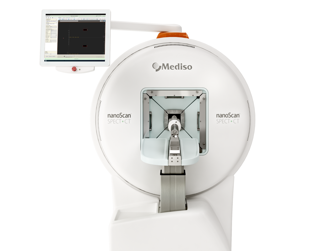
The 111In-labeled-liposome or the 111In-labeled cyclic RGDfK-liposome was injected intravenously into the mice with human melanoma cells. They were scanned with nanoSPECT/CT PLUS 24h later. Results show that mice receiving the 111Inlabeled cyclic RGDfK-liposome showed high and specific tumor uptake. Based upon these findings, the cyclic RGDfK- liposome is said to be a promising agent for tumor imaging. Present study also reports successful design of modifying the surface of conventional liposome to add the cyclic RGDfK peptide for the targeted delivery of the radioactive cyclic RGDfK liposome to human melanoma cells, where the αVβ3 integrin was expressed.
Results from nanoSPECT/CT PLUS
Nude mice were injected subcutaneously with 2x10⁵ A375.S2 human melanoma cells in their necks. Two weeks after the injection, animals had developed nodules of similar size, about 2 mm in diameter. For taking nuclear images with NanoSPECT/CT®plus scanner system, mice were injected with the 111In-labeled liposomes (50µCi), and the images of mice was captured 24 h after an injection of radioactive liposomes.
Results show that:
- In contrast to the mice receiving the 111In-labeled liposome, mice receiving the 111Inlabeled cyclic RGDfK-liposome showed a clear nuclear image of tumor nodules due to the accumulation of the 111In-labeled cyclic RGDfK-liposome.
- Specificity of the 111In-labeled cyclic RGDfK-liposome of being targeted at tumors was confirmed by mice receiving the cyclic RGDfV peptide (1mg/kg) before having an injection of the 111In-labeled cyclic RGDfK-liposome, the data showed, when compared to the mice without receiving the cyclic RGDfV peptide, that the radioactive signal within tumors in mice receiving the same peptide did decrease.
- Additionally, the tumor-to-background ratio in the mice with an injection of the 111In-labeled cyclic RGDfK-liposome was significantly higher than the ratio in the mice with the 111In-labeled liposome. The tumor-to-background ratio in mice with an injection of the 111In-labeled cyclic RGDfK-liposome but with the cyclic RGDfV peptide treatment was, however, significantly lower than the ratio in the mice with the same injection but without the cyclic RGDfV peptide.

Full article on mdpi.com
Comment pouvons-nous vous aider?
N'hésitez pas à nous contacter pour obtenir des informations techniques ou à propos de nos produits et services.
Contactez-nous
