Blood-triggered generation of platinum nanoparticle functions as an anti-cancer agent
2020.01.28.
Xin Zeng et al., Nature Communications, 2020
Summary
Despite the large amount of research on the effects of metal nanoparticles (NPs) in nature and medicine, there has been very limited application in the clinic due to their potential toxicity, cost, and ethical hurdles of research in humans.
In this Nature Communications article, the authors have discovered that platinum (Pt) nanoparticles (NPs) are generated in vivo in human blood when a patient is treated with cisplatin, a powerful anti-cancer agent. They have shown that the self-assembled Pt NPs form rapidly, accumulate in tumors, and remain in the body for an extended period. Furthermore, the Pt NPs by themselves act as anti-cancer agent, but the tumor inhibitory activity is greatly increased when the nanoparticles are loaded with a chemotherapeutic drug, daunorubicin (DNR). The Daunorubicin loaded nanoparticles appeared to be effective even in daunorubicin-resistant models.
The authors proposed that in vivo-generated metal NPs represent a biocompatible drug delivery platform for chemotherapy resistant tumor treatment.
Results from nanoScan SPECT/CT
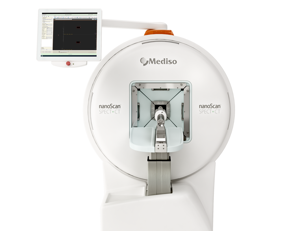
Authors have used nanoScan SPECT/CT to create high resolution images to track the tumor targeting dynamics of the nanoparticles in vivo.
- Human-derived Pt NPs were labeled with 125-I and 500 μCi 125I-Pt NPs was directly injected into DNR-resistant K562-xenografted nude mice. The images were acquired for 30 min at 1, 4, 24 and 48 h time point.
- The radioactive signal accumulated in the tumor regions, peaking at 24 h and remaining apparent at 48 h P.I. (Fig. 4f), indicating that the Pt NPs were efficiently taken up by the tumors.

Figure 4. f NanoScan SPECT/CT imaging of 125I-Pt NPs in DNR-resistant K562 cell-xenografted nude mice (n = 5) at 1, 4, 24 and 48 h after intravenous injection of the NPs. The arrows and dotted circles indicate the tumors. MIP: Maximum Intensity Projection.
Read the full article in Nature Communications
Comment pouvons-nous vous aider?
N'hésitez pas à nous contacter pour obtenir des informations techniques ou à propos de nos produits et services.
Contactez-nous
