Towards Optimized Bioavailability of 99mTc Labeled Barbiturates for Non invasive Imaging of Matrix Metalloproteinase Activity
2022.06.09.
Lisa Honold et al, Molecular Imaging and Biology, 2022
Summary
Introduction: Dysregulated activity of matrix metalloproteinases (MMPs) drives a variety of pathophysiological conditions. Non-invasive imaging of MMP activity in vivo promises diagnostic and prognostic value. However, current targeting strategies by small molecules are typically limited with respect to the bioavailability of the labeled MMP binders in vivo. To this end, we here introduce and compare three chemical modifcations of a recently developed barbiturate-based radiotracer with respect to bioavailability and potential to image MMP activity in vivo.
Methods: Barbiturate-based MMP inhibitors with an identical targeting unit but varying hydrophilicity were synthesized, labeled with technetium-99m, and evaluated in vitro and in vivo. Biodistribution and radiotracer elimination were determined in C57/BL6 mice by serial SPECT imaging. MMP activity was imaged in a MMP-positive subcutaneous xenograft model of human K1 papillary thyroid tumors. In vivo data were validated by scintillation counting, autoradiography, and MMP immunohistochemistry.
Results: We prepared three new 99mTc‐labeled MMP inhibitors, bearing either a glycine ([99mTc]MEA39), lysine ([99mTc]MEA61), or the ligand HYNIC with the ionic co-ligand TPPTS ([99mTc]MEA223) yielding gradually increasing hydrophilicity. [99mTc]MEA39 and [99mTc]MEA61 were rapidly eliminated via hepatobiliary pathways. In contrast, [99mTc]MEA223 showed delayed in vivo clearance and primary renal elimination. In a thyroid tumor xenograft model, only [99mTc]MEA223 exhibited a high tumor- o-blood ratio that could easily be delineated in SPECT images.
Conclusion: Introduction of HYNIC/TPPTS into the barbiturate lead structure ([99mTc]MEA223) results in delayed renal elimination and allows non-invasive MMP imaging with high signal-to-noise ratios in a papillary thyroid tumor xenograft model.
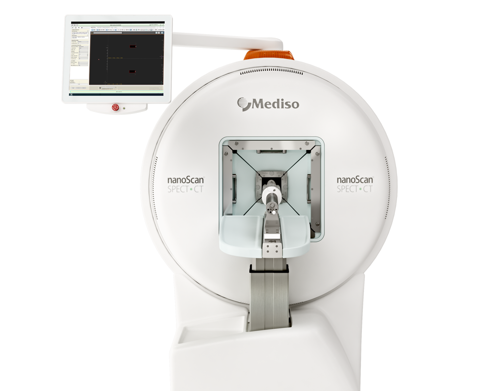
Results from nanoScan® SPECT/CT
In vivo biodistribution was determined in adult female C57/BL6 mice. Dynamic SPECT scans were acquired over the course of 90 min p.i. (9 × 10 min frames, field of view 108 mm). Following the acquisition, CT contrast agent (Ultravist®-370, 5 µl/g bw) was injected via the tail vein catheter, and a CT image was obtained. Mice underwent subsequent SPECT/ CT scans 4 h p.i. (1 × 30 min frame) and 24 h p.i. (1 × 60 min frame). For in vivo tumor uptake studies, mice were imaged 0–60 min and 4 h p.i. of tracer with a reduced feld of view (1 × 30 min frame, 26 mm).
For tumor studies, 2 × 10⁶ K1-LITG human thyroid cancer cells in 40–60 µl plain DMEM medium were subcutaneously injected above each shoulder of CD1nude/nude mice. Imaging experiments were performed 15 days post-implantation.
Results show that:
- [99mTc]MEA39 presented with a fast clearance from the blood accumulating in the excretion organs liver and kidney already within the frst 10 min. After 90 min p.i., 96.4 ± 2.0%ID were excreted, the vast majority via the hepatobiliary system.
- Elimination of [99mTc]MEA61 was equally fast, showing 93.0 ± 3.6%ID accumulating in excretion organs after 90 min p.i.. Similar to [99mTc]MEA39, the majority of [99mTc]MEA61 (92.0%) was eliminated via the hepatobiliary pathway.
- In contrast, [99mTc]MEA223 showed a delayed elimination with only 66.6 ± 4.4%ID found in excretion organs 90 min p.i.. First pass effect and early accumulation in the liver was signifcantly reduced ([%ID/ml] at 10 min p.i.: 7.3 ± 0.5 vs. 38.1 ± 9.3 ([99mTc]MEA61) vs. 50.8 ± 9.3 ([99mTc]MEA39) (Fig. 1C), and elimination was shifted towards renal elimination (64.4 ± 9.3%). Until 60 min p.i., [99mTc]MEA223 showed signifcantly higher radioactivity in the blood than [99mTc]MEA39 and [99mTc]MEA61 (Fig. 1B).

Fig. 1
- Late time point acquisitions were performed 4h and 24h after tracer injection to assess delayed kinetics: 4h p.i. blood radioactivity of [99mTc]MEA223 and [99mTc]MEA61 remained considerably high, while [99mTc]MEA39 showed an increased wash out and lower blood concentrations as shown in Fig. 2B and C. While the blood radioactivity of [99mTc]MEA223 decreased further over the next 20 h, [99mTc]MEA61 presented with a long circulation time and unchanged blood radioactivity. Radiotracer accumulation in non-excretion organs 4 and 24 h p.i. was very low for all compounds with < 1% ID/ml, but, due to increased circulation times, as a tendency slightly higher for [99mTc]MEA61.
- Complementary ex vivo measurements of tissue samples at 25 h p.i. confrmed the in vivo data at the latest time point (24 h p.i.) as shown in Fig. 2D.

Fig. 2
- Three radiotracers were applied for in vivo imaging of MMP activity in a subcutaneous xenograft model of human K1 papillary thyroid tumors which are known for high MMP activity. MMP-2 and MMP-9 activity in patients with papillary thyroid tumors are linked to tumor cell invasion and metastasis, and high gelatinase activity has been associated with poor prognosis
- 1 h p.i., in vivo tumor signal was highest for [99mTc]MEA223, followed by [99mTc]MEA61 and [99mTc]MEA39. Diferences in early tumor signals were in accordance with diferences in blood radioactivity, as tumor-to-blood ratios remained below 1 for all radiotracers. 4 h p.i., [99mTc]MEA223 showed a strong accumulation in tumors, independent of local tumor perfusion with positive tumor-to-blood ratios. In contrast, [99mTc]MEA61 and [99mTc]MEA39 showed no specifc tumor accumulation, as tumor signals declined parallel to the decrease in blood radioactivity. In vivo results could be confrmed by ex vivo scintillation counting. Additionally, ex vivo autoradiography confrmed that only [99mTc]MEA223 showed relevant radiotracer accumulation throughout the tumor, even though all tumors were highly positive for the target in MMP immunohistochemistry.

Fig. 3
Tu sum up: amino acid-based MMP tracers [99mTc]MEA39 and [99mTc]MEA61 were rapidly eliminated via hepatobiliary pathways. In contrast, the more hydrophilic [99mTc]MEA223 was primarily excreted via the kidneys and showed a signifcantly increased bioavailability for the frst 90 min after injection. In a thyroid tumor xenograft model [99mTc]MEA223 exhibited a high tumor-to-blood ratio that could easily be delineated in SPECT images. The newly developed [99mTc]MEA223 hence allows non-invasive imaging of MMP activity with high signal-to-noise and should be investigated in additional pathophysiological conditions.
Full article on link.springer.com
Comment pouvons-nous vous aider?
N'hésitez pas à nous contacter pour obtenir des informations techniques ou à propos de nos produits et services.
Contactez-nous
