GMP production of [68Ga]Ga-BOT5035 for imaging of liver fibrosis in microdosing phase 0 study
2020.07.28.
Irina Velikyan et al, Nuclear Medicine and Biology, 2020
Summary
Introduction: Early detection of liver fibrosis and monitoring response to treatment crucial for the management of patients are currently not feasible in clinical practice. Platelet derived growth factor receptor β (PDGFR-β) expression is regarded as a potential biomarker to determine the stages of fibrotic diseases including liver fibrosis. [68Ga]Ga-BOT5035 comprising a bicyclic peptide was developed for specific targeting of PDGFR-β overexpressed in pathological fibrosis. The realization of microdosing phase 0 study using [68Ga]Ga-BOT5035 positron emission tomography required automated good manufacturing practice (GMP) compliant production of [68Ga]Ga-BOT5035 presented herein. Moreover, the investigation of radiation dosimetry was conducted to ensure possibility of multiple annual examinations for disease monitoring in clinical setup.
Methods: The active pharmaceutical ingredient starting material BOT5035 was provided by BiOrion Technologies BV. The 68Ga-labelling process was developed and automated using synthesis platform, disposable cassettes for 68Ga-labelling, and pharmaceutical grade 68Ge/68Ga generator. Radiolysis sensitive BOT5035 required development and systematic optimization of the labelling synthesis parameters. The validation process was conducted with regard to the product quality and quantity, as well as production reproducibility. The kinetics of the uptake of [68Ga]Ga-BOT5035 in organs of interest was evaluated with nanoScan PET/MRI 3T in mice. Human organ equivalent doses and total body effective doses were calculated based on ex vivo organ distribution in Sprague-Dawley rats.
Results: The GMP compliant automated production of [68Ga]Ga-BOT5035 with on-line documentation demonstrated high reproducibility. The time for the labelling synthesis and quality control was approximately 60 min. Ex vivo organ distribution data revealed fast blood clearance and washout from most of the organs. The dose-limiting organs were kidney and bone marrow. The total effective dose as limiting parameter would allow for up to 3–4 PET scans per annum.
Conclusion: The fully automated and GMP compliant production of [68Ga]Ga-BOT5035 was developed and thoroughly validated. The radiopharmaceutical was approved by Swedish Medicinal Products Agency and the Ethical Review Authority for the Phase 0 clinical study of the quantitative imaging of liver fibrosis. Human dosimetry calculations extrapolated from animal experiment indicated possibility of 3–4 PET examinations per year.
Results from nanoScan PET/MRI 3T and nanoScan SPECT/CT
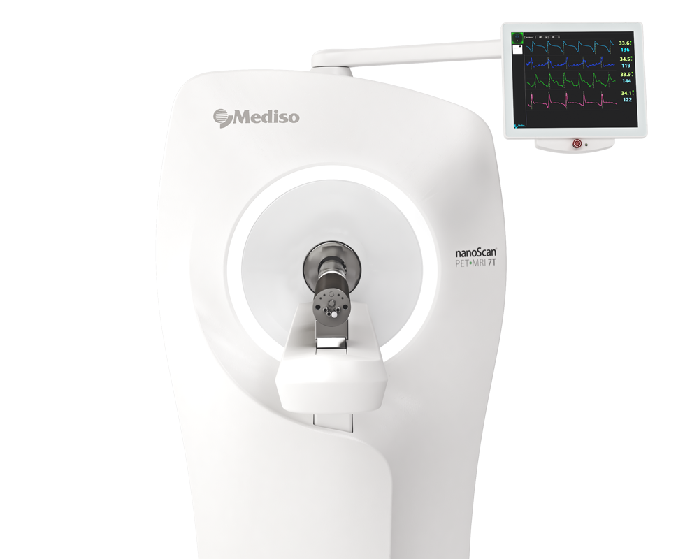
BALB/c mice were anaesthetized by sevoflurane gas inhalation. An i.v. catheter was placed in the lateral tail vein. PET images were acquired in a nanoScan PET/MRI 3T animal scanner
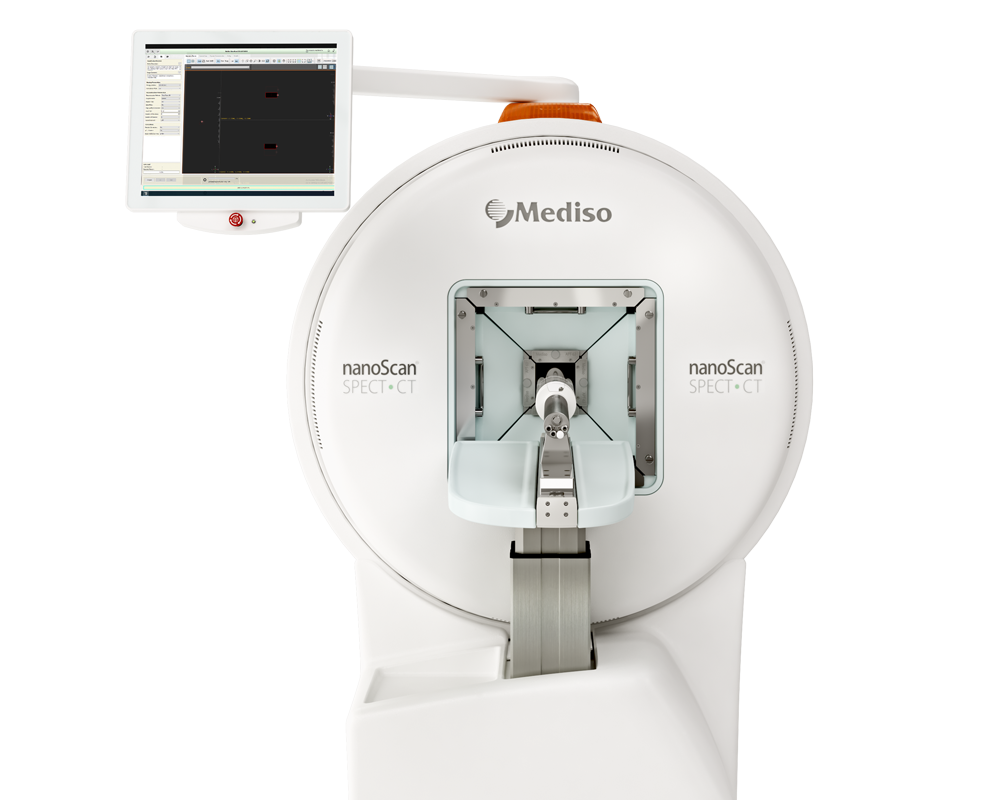
followed by a CT examination in nanoScan SPECT/CT. Following imaging, the rodents were euthanized. Organs were harvested and weighed, and an assessment of radioactivity was performed. The kinetics of the uptake of [68Ga]Ga-BOT5035 in organs of interest was evaluated with PET/MRI. A total of 2 female mice were administered 0.7–5.0MBq of [68Ga]GaBOT5035 through the catheter placed in the lateral tail vein. A dynamic PET-scan was performed for 60 min, using the abdomen as field of view. Whole body CT scan was performed for 8 min. PET images were reconstructed using a TeraTomo 3D algorithm with 4 iterations and 6 subsets. PET/CT images were fused and analysed in PMOD. Fused PET/CT images were presented as maximum intensity projection (A).
Results show:
- The dynamic scanning revealed fast clearance from healthy organs except from kidneys. The blood clearance kinetics was best described by a two-phase exponential decay (C). The accumulation in the kidneys reached a plateau at 60 min (B). Uptake in the urinary bladder continued to increase after 30 min indicating excretion. The wash out from the liver was fast (B).
 Full article on sciencedirect.com
How can we help you?
Don't hesitate to contact us for technical information or to find out more about our products and services.
Get in touch
