99mTc-NTP 15-5 is a companion radiotracer for assessing joint functional response to sprifermin (rhFGF-18) in a murine osteoarthritis model
2022.05.17.
Arnaud Briat et al., 2022, Scientific Reports
Summary
Osteoarthritis (OA) is a slowly, progressive, ultimately degenerative disorder of movable joints, mainly identified at the clinical level by pain and functional limitation and involving all joint structures. OA progression can be characterized by joint cartilage degradation and loss, subchondral bone remodelling, and synovial membrane inflammation. Current treatments can improve symptoms but do not delay the progression of disease, the total joint replacement finally becoming the only possible outcome for many patients.
The articular cartilage is responsible for the biomechanical properties of the joint, supported by an extracellular matrix (ECM), rich in fibrillar proteins such as collagens, and in proteoglycans (PG). PG contain covalently bound glycosaminoglycans (GAGs), which are essential to the function of the molecule as they draw water into the cartilage matrix, giving it the ability to withstand compression. During OA development, a loss of PG and a disruption of the collagen network occur, leading to the matrix destruction and ultimately to the total cartilage loss. Therefore, the articular cartilage and its ECM appear as therapeutic targets of importance.
With the better understanding of the pathophysiology of OA progression, promising therapeutic targets have been identified, with the emergence of Disease Modifying OsteoArthritis Drugs (DMOAD), aiming at not only assisting with symptom management but also modifying the structural course of the disease. Despite extensive research on DMOAD, there is currently no pharmacological intervention approved for use in Europe or the USA demonstrating efficacy for modifying OA progression. Even if promising DMOADs have emerged, the major challenge in demonstrating the proof of concept is to overcome (i) the absence of a precise assessment of the disease, particularly in the early stages, and (ii) the lack of consensus on how to detect joint changes and link them to clinically meaningful endpoints. Therefore, new treatments are needed, as well as new methods to evaluate their efficacy and to monitor OA progression. Authors indeed consider that research and development towards DMOADs is hampered by the lack of specific and sensitive imaging tools to reliably quantify OA progression, and monitor response to therapy, including radiographs and MRI. Higher sensitive imaging approaches appear of real need to evaluate OA progression and to monitor response to innovative therapies.
Few DMOAD currently under development are aiming at preventing cartilage deterioration and/or restoring cartilage thickness. Among them, sprifermin, a recombinant human Fibroblast Growth Factor 18 (rhFGF-18), appeared as the most promising molecule. In rats and in human explants, it has been demonstrated that FGF-18 increased chondrocytes proliferation, as well as ECM deposition and PG production, leading to an increase in cartilage thickness. The strong anabolic effect of sprifermin seems to follow a sequential process, with an early increase of aggrecanase activity leading to aggrecan degradation, described as a prerequisite for cell proliferation, followed by ECM molecules production by newly produced chondrocytes, including sustained PG production, and ultimately cartilage regeneration. To date, sprifermin is the only candidate DMOAD with a proven structural effect on cartilage. The clinical relevance of the results obtained is currently being discussed. This makes it the only possible gold standard to date for a study to investigate a new surrogate marker of cartilage remodelling.
In such a context, the authors' group aims at validating a nuclear medicine imaging strategy targeting cartilage PG in vivo, which could provide a functional access to joint and a new method to evaluate innovative therapies. Because cartilage contains up to 10% PG consisting of mainly chondroitin sulphate aggrecan which chains are negatively charged, their strategy is based on the use of a bi-functional agent, the radiotracer 99mTc-N-(triethylammonium)-3-propyl-[15]ane-N5 (99mTc-NTP 15-5), that contains in its structure a positively charged quaternary ammonium function for binding to PG and a polyazamacrocycle to complex 99mTc. Based on many preclinical studies, the authors believe that 99mTc-NTP 15-5 and in vivo functional imaging of PG could provide a suitable set of criteria for quantifying cartilage functionality, and the efficacy of new emerging therapeutic strategies.
In this study, the authors determined the relevance and sensitivity of 99mTc-NTP 15-5 imaging in the destabilization of the medial meniscus osteoarthritis murine model, for assessing the structural effect of sprifermin, with the opportunity to bridge the gap between preclinical and clinical testing.
Results from the nanoScan SPECT/CT
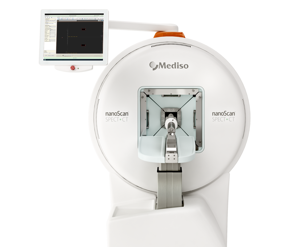
SPECT-CT imaging was performed 30 min after the intravenous injection of 20 MBq of 99mTc-NTP 15-5. Multimodal SPECT-CT imaging was performed using the NanoScan SPECT-CT camera equipped with four detectors and multi pinhole collimation (APT62). Mice were anesthetized with isoflurane (4% for induction, and 1.5% during images acquisition) and placed in a Multicell Mouse L bed with temperature control (37 °C). Nucline software (Nucline 3.00.018, Mediso Ltd) was used for image acquisitions and reconstructions. CT parameters: helical scan with 480 projections (300 ms per projection), 50 kV, 590 μA, pitch 1.0, binning 1:4 and field of view: max. SPECT acquisitions parameters: images were acquired within the CT scan range, with resolution set to “standard”. The time per projection was determined in accordance with the detected radioactivity (most frequently used: 30 s). SPECT images reconstruction were conducted using TeraTomo3D (Nucline v3.00.018, Mediso Ltd) with a normal dynamic range. Regularization filters, reconstruction resolution and iterations were set to “medium”. Additional corrections were performed during reconstruction: Monte Carlo correction quality was set to “high”; Attenuation: based on CT attenuation map and scatter corrections; Activity decay correction: during acquisition time lapse.
99mTc-NTP 15-5 activity was quantified in a volume of interest delineated over the operated (pathologic) and non-operated (contralateral) knees. The activity ratios between pathologic over contralateral knees were calculated for each animal at each time point.
Figure 1. shows in vivo distribution by SPECT-CT imaging of 99mTc-NTP 15-5 30 min after intravenous injection of 20 MBq of radiotracer in healthy mice. (A) Representative transversal, coronal, and sagittal slices of 99mTc-NTP 15-5 distribution in anesthetized mice. From left to right: transversal views at the humeral head level (dotted line 1) and at the knees level (dotted line 2); coronal and sagittal whole-body views. Arrows indicate cartilage uptake in the mouse shoulders, knees, and intervertebral disks, respectively. Non-specific uptake is observed in the liver (L) and bladder (B). SPECT scaling range is 65 to 650 kBq/ml. (B) Representative SPECT-CT Maximum Intensity Projection 3D image focused on knees. Specific uptake in femoral condyle (FC) and tibial plateau (TP) is observed, as well as non-specific accumulation in bladder (B). SPECT scaling range is 650 to 2000 kBq/ml.
- The authors' experimental results in the DMM model of OA treated by sprifermin demonstrate the proof of concept that 99mTc-NTP 15-5 imaging could provide an early and sensitive tool to evaluate cartilage remodelling and DMOAD response.
- These promising results are in favour of reinforcing first into humans transfer for evaluating the sensitivity of 99mTc-NTP 15-5 imaging in patients with OA, that the authors have recently initiated (NCT04481230).
How can we help you?
Don't hesitate to contact us for technical information or to find out more about our products and services.
Get in touch
