Features & Benefits
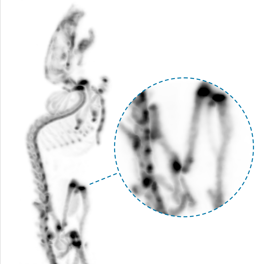
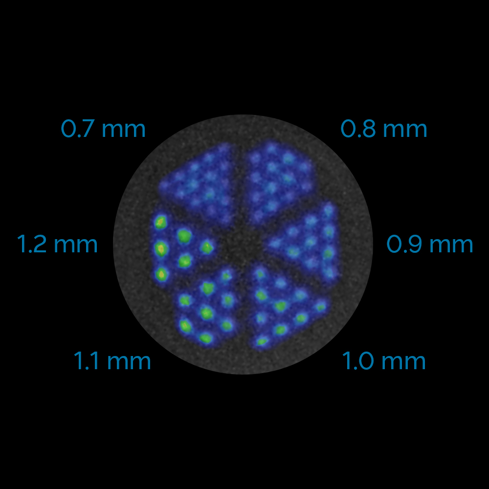
Resolving precise details with 700 μm spatial resolution
- Finest (1.12mm×1.12mm) lutetium oxyorthosilicate (LSO) crystal needles provide precise signal localization preserving spatial information in raw data
- Tera-Tomo™ 3D PET iterative reconstruction with real-time Monte Carlo based physical modelling unveiling the tiniest details on the image
- Large ring diameter and statistical depth of interaction compensation offers homogeneous image quality over the entire field of view
Uncompromised applications with very low level of radioactivity
- Thick LSO crystals for excellent sensitivity
- Short (3 ns) coincidence time window necessary for improved signal to noise ratio
- Advanced corrections (random, scatter, LSO background etc.) ensuring quantification at low activity levels
- Best Minimal Detectable Activity on the market: 60 Bq (1.6 nCi)
- Dedicated feature of the iterative Tera-Tomo™ 3D PET reconstruction engine ensures precise quantification at very low levels of activity
- Analytic reconstruction option (FBP) with attenuation and scatter correction available for quantitative reconstruction of low activities close to regions with significantly higher activity
- Inherently optimized for longitudinal studies e.g. long-term cell tracking.
Coping with count rate: mastering studies with high dose
- Multichannel read-out electronics with ultra-fast data processing and advanced dead-time correction
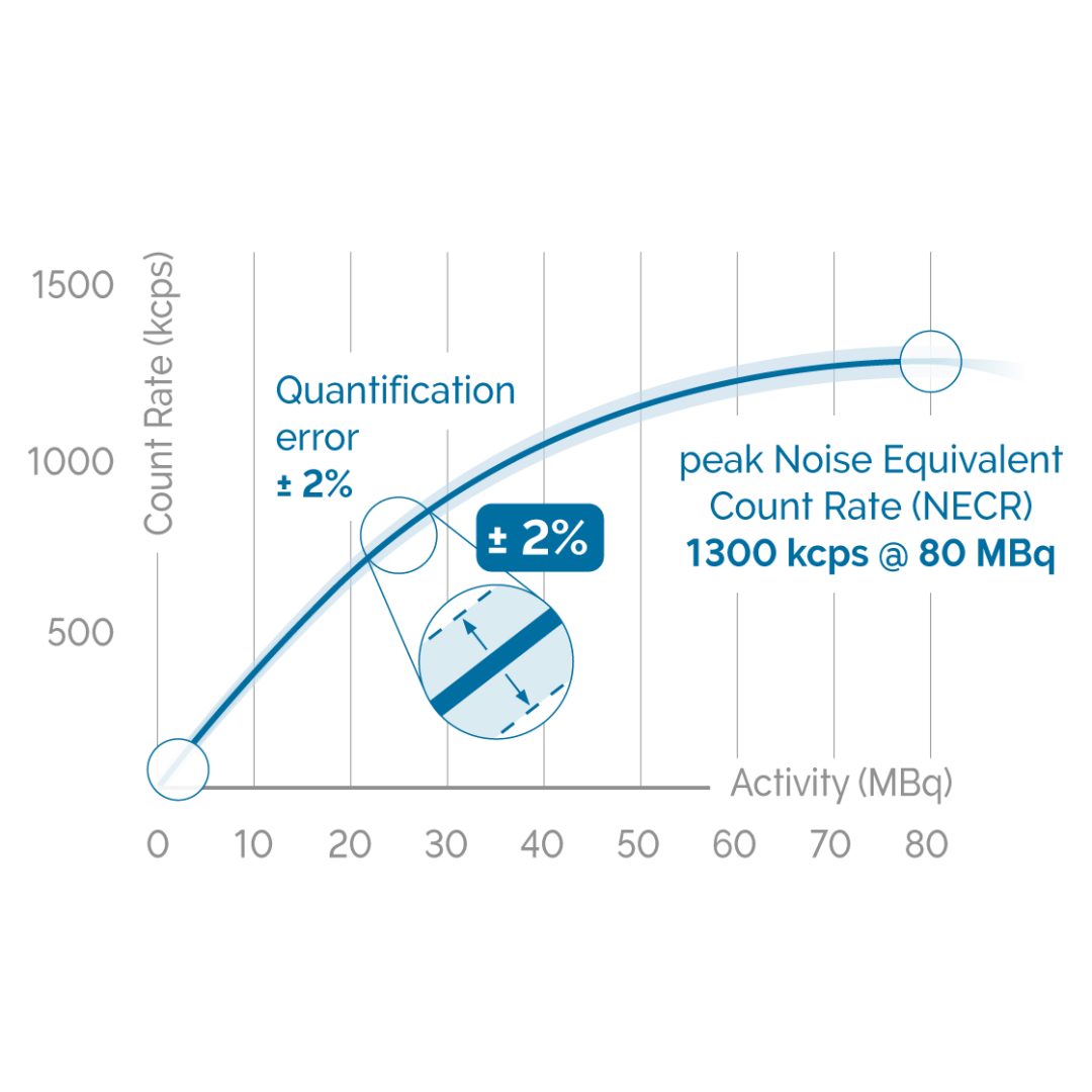
- Exceptional count rate performance – peak Noise Equivalent Count Rate (NECR) for mouse is up to 1300 kcps @ 80 MBq (2.16 mCi)
- Accurate and reproducible uptake values ensured by very high level of quantification accuracy up to 80 MBq (2.16 mCi) and beyond, with quantification error of ±2%
- Suitable for dynamic imaging of large animals or for dynamic scanning of up to four mice or two rats simultaneously
- Optimal for imaging of isotopes with short half-life (11C, 13N, 15O, etc.)
Largest transaxial field of view
- Wide bore diameter up to 16 cm allowing free access to the animals
- Large transaxial field of view up to 12 cm
- Excellent homogeneity over the entire field of view
- Suitable for various animal models from tiny mouse (25 g) to large rabbits (6.5 kg)
- Simultaneous multiple animal imaging (up to 4 mice or 2 rats) with individual physiological monitoring
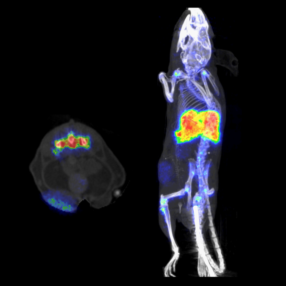
Excellent image quality and quantitation accuracy with the Tera-Tomo™ image reconstruction
Our proprietary iterative reconstruction engine, used in both clinical and preclinical systems ensures quantitative results with excellent resolution for all PET isotopes.
- The Tera-Tomo™ 3D PET reconstruction engine along with the most advanced detector calibration algorithms ensure a very high level of quantification accuracy through the entire dynamic range of the system. The quantification error of +/- 2% results in accurate and reproducible uptake values
- 3D iterative reconstruction applying deep Monte Carlo based physical modelling of particle-level interaction from positron emission to detection
- Advanced corrections for energy, time, dead-time and random events and positron range
- CT or MR based attenuation and scatter correction
- Automated workflow with 4D multiparametric PET reconstruction for fast kinetic analysis of dynamic acquisitions
High power CT with large field-of-view and high resolution
- Up to 80 W X-ray power together with the largest field of views (12 cm transaxial and 45 cm helical scan range) enables high performance scanning of even large or multiple animals
- Very high throughput: fast scanning with real-time image reconstruction combined with multiple animal imaging capability
- Variable magnification (up to x7.6) for high-resolution imaging even with 10 μm isotropic voxel size
- Iterative image reconstruction for low-noise and low-dose imaging
- Ultra-low-dose protocol for follow-up studies with less than 1 mGy whole-body dose to the animal for reliable longitudinal studies, e.g. following tumour growth or therapy response
- Imaging with cardiac and respiratory gating for reducing motion artifacts and for analysis of cardiac and pulmonary function.
Applications
Publications
Images
Specification
up to 16 cm
up to 12 cm
up to 15 cm
mouse, rat, marmoset, guinea pig, rabbit
up to 4×60g mice 2×300g rats
LSO (1.12×1.12×13 mm)
down to 0.7 mm
down to 1.25 mm
up to 10.5% (250-750 keV)
up to 1800 kcps @ 90 MBq / 2.43 mCi
up to 850 kcps @ 70 MBq / 1.89 mCi
up to 80 W
16 cm
12 cm
10 cm
mouse, rat, marmoset, guinea pig, rabbit
up to 4×60 g mice 2×300 g rats
modified Feldkamp-type for real-time reconstruction, iterative for low-dose and low-noise applications
30 μm at 10 μm voxel size
down to 1 mGy for whole-body mouse
Maintenance-free permanent magnet with 1 Tesla field strength
Zero (5 Gauss line is inside the cover)
Integrated RF shielding, no need for Faraday cage
Volume coils: whole-body mouse, whole-body rat; Surface coils: mouse brain, rat brain
100 μm in vivo <100 µm ex vivo
Downloads
How can we help you?
Don't hesitate to contact us for technical information or to find out more about our products and services.
Get in touch