SPECT/CT imaging, biodistribution and radiation dosimetry of a 177Lu-DOTA-integrin αvβ6 cystine knot peptide in a pancreatic cancer xenograft model
2021.05.31.
Sachindra Sachindra et al, Frontiers in Oncology, 2021
Summary
Introduction: Pancreatic ductal adenocarcinoma (PDAC) is one of the most aggressive malignant neoplasms, as many cases go undetected until they reach an advanced stage. Integrin αvβ6 is a cell surface receptor overexpressed in PDAC. Consequently, it may serve as a target for the development of probes for imaging diagnosis and radioligand therapy. Engineered cystine knottin peptides specific for integrin αvβ6 have recently been developed showing high affinity and stability. This study aimed to evaluate an integrin αvβ6-specific knottin molecular probe containing the therapeutic radionuclide 177Lu for targeting of PDAC.
Methods: The expression of integrin αvβ6 in PDAC cell lines BxPC-3 and Capan-2 was analyzed using RT-qPCR and immunofluorescence. In vitro competition and saturation radioligand binding assays were performed to calculate the binding affinity of the DOTA-coupled tracer loaded with and without lutetium to BxPC-3 and Capan-2 cell lines as well as the maximum number of binding sites in these cell lines. To evaluate tracer accumulation in the tumor and organs, SPECT/CT, biodistribution and dosimetry projections were carried out using a Capan-2 xenograft tumor mouse model.
Results: RT-qPCR and immunofluorescence results showed high expression of integrin αvβ6 in BxPC-3 and Capan-2 cells. A competition binding assay revealed high affinity of the tracer with IC50 values of 1.69 nM and 9.46 nM for BxPC-3 and Capan-2, respectively. SPECT/CT and biodistribution analysis of the conjugate 177Lu-DOTA-integrin αvβ6 knottin demonstrated accumulation in Capan-2 xenograft tumors (3.13 ± 0.63%IA/g at day 1 post injection) with kidney uptake at 19.2 ± 2.5 %IA/g, declining much more rapidly than in tumors.
Conclusion: 177Lu-DOTA-integrin αvβ6 knottin was found to be a high-affinity tracer for PDAC tumors with considerable tumor accumulation and moderate, rapidly declining kidney uptake. These promising results warrant a preclinical treatment study to establish therapeutic efficacy.
Results from nanoScan® SPECT/CT plus
For in vivo experiments, at least 8-week-old female athymic NMRI-Foxn1nu/Foxn1nu mice were used. For the generation of tumor xenografts, 5 x 10⁶ cells of Capan-2 cells were inoculated subcutaneously into the left and right shoulder. Tumors were allowed to grow for two to four weeks (tumor volume > 100 mm3).
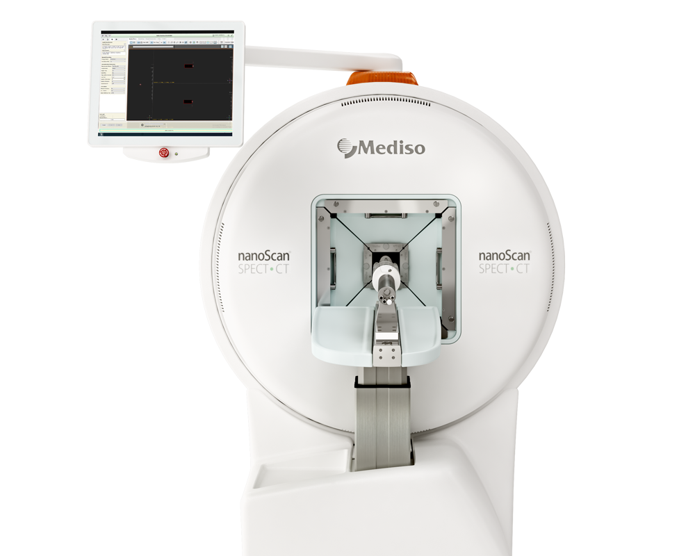
SPECT and CT imaging were performed using the nanoSPECT/CTplus scanner. Mice were anesthetized using 1-2% isoflurane with oxygen at a flow rate of approximately 0.5 l/min. After a low-dose CT scan for positioning the tumors and kidneys in the scan range the SPECT acquisition was started directly before intravenous injection of approximately 50 MBq of 177Lu-DOTA-integrin αvβ6 knottin (0.1-0.15 ml). Nine consecutive multi-pinhole SPECT images of 10 min duration each (5 angular steps a 60 sec, 2 bed positions) were acquired. Additional individual scans of 35-60 min duration (5 angular steps á60-180 sec, 2 bed positions) were performed up to 8 days to assess biodistribution kinetics.
For biodistribution studies, tumor-bearing mice (n=4-5 per time point) were injected with approximately 1 MBq (22 pmol) of 177Lu-DOTAintegrin αvβ6 knottin to the tail vein via a catheter. Mice (n=4-5 per time point) were sacrificed and dissected one, two, three and eight days post injection. Tumor, blood, stomach, pancreas, small intestine, colon, liver, spleen, kidney, heart, lung, muscle and femur samples were weighed and uptake of radioactivity was measured by a gamma counter.
SPECT/CT imaging revealed:
- accumulation of 177Lu-DOTA-integrin αvβ6 knottin in both the tumors and the kidneys (Figure 3A)
- uptake by other organs was low or moderate
- short- and long-term tracer kinetics for kidney and tumor were quantified from SPECT images, up to 3 and 187 hours post-injection: kidneys showed a higher initial accumulation of 177Lu-DOTA-integrin αvβ6 knottin (Figure 3B), yet clearance was also faster than from the tumor (Figure 3C)

FIGURE 3 | SPECT/CT imaging with 177Lu-DOTA-integrin αvβ6 knottin. (A) SPECT images show maximum intensity projection (MIP), coronal and transverse projections fused with CT images of 177Lu-DOTA-integrin αvβ6 knottin in a Capan-2 xenograft model at 22 hours post injection of 62 MBq tracer. Nude mice were carrying xenografts on left and right shoulder. (B) Early SPECT kinetics data show the uptake of 177Lu-DOTA-integrin αvβ6 knottin in tumor and kidney. (C) SPECT-based time-activity curve (2-187 hours p.i.) shows faster clearance of 177Lu-DOTA-integrin αvβ6 knottin from kidney than from tumor.
Taken together with the biodistribution analysis, in this study, binding of the peptide tracer 177Lu-DOTA-integrin αvβ6 knottin to its target both in vitro and in vivo was investigated. 177Lu-DOTA-integrin αvβ6 knottin exhibited high affinity and specific binding to target-positive cells and tumors. The study demonstrated the translational potential of this tracer for imaging and therapy of integrin αvβ6-overexpressing tumors like PDAC.
Full article on frontiersin.org
W czym możemy pomóc?
Skontaktuj się z nami aby uzyskać informacje techniczne i / lub wsparcie dotyczące naszych produktów i usług.
Napisz do nas
