SPECT imaging of distribution and retention of a brain-penetrating bispecific amyloid-β antibody in a mouse model of Alzheimer’s disease
2020.09.21.
Tobias Gustavsson et al., Translational Neurodegeneration, 2020
Summary
Currently, approximately 45 million people worldwide are affected by Alzheimer’s disease (AD). To date, there is no treatment that can halt the neurodegenerative processes of AD. Biopharmaceutical drugs, such as monoclonal antibodies (mAbs), have advantages of high affinity and target specificity combined with the ability to initiate multiple different downstream effector functions, facilitating treatment effects.
Intracellular neurofibrillary tangles of hyperphosphorylated tau and extracellular plaques composed of aggregated amyloid-β (Aβ) are pathological hallmarks of AD. Although large insoluble Aβ fibrils are the main constituent of plaques, the smaller soluble oligomeric and protofibrillar Aβ species have been suggested to be linked to disease progression and impaired synaptic function, and may thus provide a better therapeutic target. BAN2401, a conformation-dependent Aβ mAb that binds selectively to soluble Aβ oligomers and protofibrils, reduced Aβ load and slowed the cognitive decline in a dose-dependent manner in a large phase 2b study of 856 prodromal and mild AD patients.
However, brain entry of large and hydrophilic molecules is severely limited by the blood-brain barrier (BBB) and the blood-cerebrospinal fluid barrier (BCSFB). Bispecific antibodies that display dual-target affinity, for example targeting an endogenous BBB transport mechanism and a brain parenchymal target, such as Aβ, have been developed to increase brain uptake. One such BBB transporting system is the transferrin receptor (TfR), which transports the iron-binding protein transferrin across the endothelium of the BBB. The TfR can also be used to deliver biological drugs across the BBB.
The authors have previously developed a bispecific mAb, RmAb158-scFv8D3, based on RmAb158 (recombinant murine version of BAN2401) that is fused to a single-chain variable fragment (scFv) of the mouse TfR mAb 8D3. RmAb158-scFv8D3 can rapidly cross the BBB and displays faster blood clearance than the unmodified RmAb158. It is unclear how the rapid blood clearance of RmAb158-scFv8D3 affects the therapeutic efficacy of this bispecific antibody in a long-term perspective.
In this study, the authors set out to longitudinally assess the brain retention and distribution of RmAb158-scFv8D3 and RmAb158 using single-photon emission computed tomography (SPECT), during a four-week period in the tg-ArcSwe mouse model of AD.
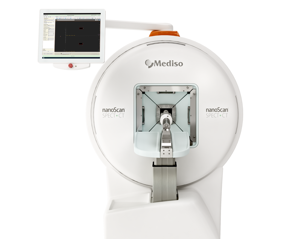
Results from the nanoScan SPECT/CT:
Mice that underwent SPECT scanning were given water supplemented with 0.2% NaI throughout the study to reduce thyroidal uptake of free 125I. Mice were intravenously (i.v.) injected with 10.39 ± 1.91 MBq [125I]RmAb158-scFv8D3 (molar activity at the time of administration: 129 ± 32 MBq/nmol; dose: 0.65 ± 0.14 mg/kg) (n = 9) or 8.67 ± 0.84 MBq [125I]RmAb158 (molar activity at the time of administration: 117 ± 62 MBq/nmol; dose: 0.25 ± 0.07 mg/kg) (n = 5). SPECT scans were obtained at 3, 6, 14, and 27 days after injection. Each mouse underwent a maximum of three SPECT scans. Mice were anesthetized with 3% sevoflurane before scanning, and then positioned on the pre-heated scanner bed of the small animal nanoScan SPECT/CT. CT was performed with the following settings: 50 kilovoltage peak X-ray, 600 μA, and 480 projections; and CT images were reconstructed using filtered back projections. 125I γ emission was collected with an acquisition frame of 2 min.
SPECT acquisition data were reconstructed using Nucline software and Tera-Tomotm 3D SPECT reconstructive algorithm with scattering and attenuation correction. SPECT images were reconstructed using 48 iterations into a static image. Reconstructed data were decay-corrected and adjusted for injected dose.
Figure 2. shows the main results from the SPECT/CT acquisitions: [125I]RmAb158-scFv8D3 displayed high brain uptake and a uniform distribution pattern in areas with known Aβ pathology. The SPECT images showed that [125I]RmAb158-scFv8D3 resided in the brain at higher concentrations than [125I]RmAb158 during the whole study period of 27 days. In addition, the intrabrain distribution of [125I]RmAb158-scFv8D3 was spatially relatively stable during the study, with signals slowly decreasing in all brain regions over time. These observations were further confirmed with ex vivo autoradiography, which demonstrated a uniform distribution pattern and decreased antibody retention over time (a).
In contrast, [125I]RmAb158 displayed a fundamentally different distribution pattern (b). At 3 days post-injection, SPECT scanning revealed no detectable [125I]RmAb158 signal in Aβ-abundant brain areas, as the measured signals were mostly blood-derived background activity. At 6 days, [125I]RmAb158 retention was visible in central brain areas in two out of three scanned mice. At 14 and 27 days post-injection, SPECT scanning showed further increased [125I]RmAb158 retention in regions that appeared to be ventricles, in both the coronal and sagittal views (b). In contrast to [125I]RmAb158-scFv8D3, ex vivo autoradiography revealed hotspots of antibody retention in the cortex and central brain (b). There was a gradual loss of antibody from the cortex, while the central brain areas displayed a more sustained antibody signal.
[125I]RmAb158-scFv8D3 displayed no brain retention in WT mice 3 days post injection (c), while [125I]RmAb158 showed a faint signal centrally in the brain (d).
- From an imaging perspective, this study demonstrated that the brain retention of unmodified and bispecific antibodies labeled with the long-lived isotope 125I can be explored in vivo with SPECT. Traditional brain imaging ligands are usually small molecular compounds, radiolabeled with the relatively short-lived carbon-11 (11C) or fluorine-18 (18F), with nanomolar affinities. These radioligands have a biological and radioactive half-life in the range of minutes to hours, whereas the 125I-labelled antibodies used in this study have a biological and radioactive half-life in the range of days to weeks, which implies the benefit of this imaging technique for long-term studies of brain uptake and retention of antibodies. With the help of long-lived radionuclide in combination with SPECT scanning, the complete dynamics of brain distribution of the two antibodies could be followed up in vivo over a period of 1 month.
- Further, although RmAb158 was found to be co-localized with Aβ in relatively high concentrations, its distribution was not representative of the overall distribution of Aβ pathology in the brain. Therefore, a system that can actively transport antibody into the brain is essential for making use of an antibody as an imaging agent for intra-parenchymal targets.

W czym możemy pomóc?
Skontaktuj się z nami aby uzyskać informacje techniczne i / lub wsparcie dotyczące naszych produktów i usług.
Napisz do nas
