Preclinical evaluation of 68Ga-labelled Pt(IV)-DFO complexes as PET-imaging agents for osteosarcoma
2026.09.03.
López-Martínez et al., European Journal of Medicinal Chemistry, 2026
Summary
The most commonly used chemotherapy agents for osteosarcoma are platinum(II)-based compounds; however, platinum(IV) prodrugs have recently gained attention as alternatives designed to overcome the dose-limiting toxicities and resistance mechanisms associated with Pt(II) complexes. In this study, Pt(IV) prodrugs were labeled with gallium-68 68Ga using deferoxamine (DFO) as a chelator to facilitate in vivo tracking via positron emission tomography (PET) imaging. Five complexes were evaluated: four functionalized Pt(IV)-DFO complexes (A–D) and one DFO-succinic acid conjugate (1). Comprehensive in vivo characterization of the PET tracers was conducted in both naïve and orthotopic osteosarcoma mouse models. Prior to administering the 68Ga-labeled Pt(IV)-DFO tracers, tumor-bearing mice underwent 18F-FDG PET scans to baseline and monitor tumor growth
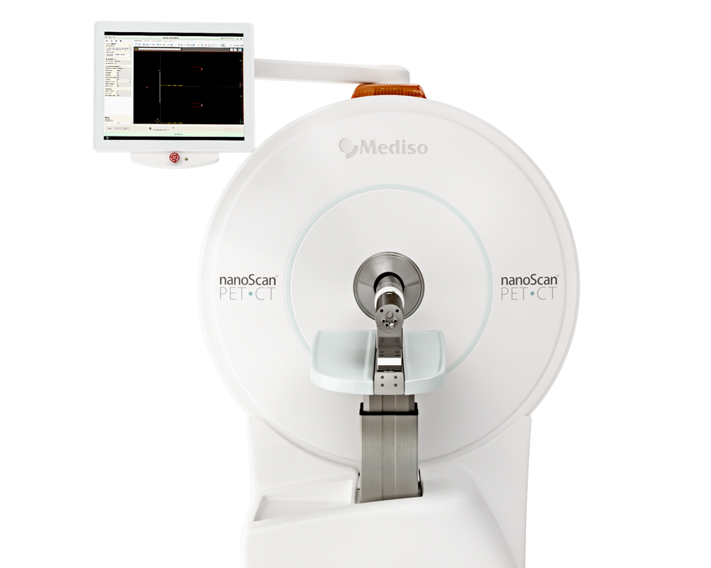
Results from nanoScan® PET/CT
Both naive (n = 12; 6 female/6 male per tracer) and osteosarcoma model mice (n = 6 per tracer) underwent dynamic PET imaging. Anesthesia was induced using isoflurane, and a 30-gauge tail-vein catheter was inserted for tracer administration. Respiration was continuously monitored, and body temperature was maintained using a heating pad set to 38 °C throughout the scan. Two animals were imaged simultaneously using a dual-bed setup per imaging session. For the 68Ga-labeled Pt(IV)-DFO measurements, 11–15 MBq in 100 μL was injected, followed by a 60-minute dynamic PET acquisition and subsequent CT imaging (720 projections, 50 kV, 980 μA, 300 ms exposure time). Tumor progression was monitored on days 7, 10, and 14 post-inoculation using 18F-FDG. For these scans, a 10-minute static PET acquisition was initiated 60 minutes post-injection, followed by low-dose CT imaging (360 projections, 50 kV, 980 μA, 170 ms exposure time).
Raw PET data were reconstructed using the Tera-TOMOTM iterative reconstruction algorithm (8 subsets, 4 iterations) with an isotropic voxel size of 0.4 mm3. Dynamic PET data were frame-binned into time windows 8 × 5 s, 2 × 10 s, 2 × 30 s, 3 × 60 s, 2 × 150 s, 2 × 300 s. CT projections were reconstructed using filtered back-projection (FBP), resulting in isotropic voxel sizes of 125 µm3 for standard CT scans and 250 µm3 for low-dose CT scans.

Fig. 1 (I) In vivo PET/CT images of female naïve mice at 15 min post-injection from all 68Ga-labelled compounds. Coronal sections show predominately renal uptake (A-E), and intestinal/hepatic distribution (F-J). The white arrows indicate different organs of interest, K: kidney; B: bladder; SI: small intestine; L: liver. (II) In vivo PET/CT images of osteosarcoma-bearing mice at 12.5 min post-injection of all different 68Ga-labelled compounds. The white arrow indicates the tumour region (T) in the left hind limb in the first image; the tumour remains in the same anatomical position across all subsequent images. (III) Area under the curve (AUC0-55min) derived from PET time-activity curved for tumour and healthy leg tissues across all 68Ga-labelled compounds. Statistical analyses were performed using a paired t-test (p < 0.05 (*), p < 0.01 (**)).
The general conclusion of this study is that the 68Ga-labeled Pt(IV)-DFO complexes performed remarkably well in the orthotopic osteosarcoma model, with [68Ga] Ga-D demonstrating the most favorable kinetic profile. Although further preclinical evaluation is required, these tracers hold significant potential for future clinical translation.
Full article on sciencedirect.com
W czym możemy pomóc?
Skontaktuj się z nami aby uzyskać informacje techniczne i / lub wsparcie dotyczące naszych produktów i usług.
Napisz do nas