Multiple‑pinhole collimators improve intra and between‑rater agreement and the certainty of the visual interpretation in dopamine transporter SPECT
2022.08.17.
Franziska Mathies et al., EJNMMI Research, 2022
Background
Multiple-pinhole (MPH) collimators improve the resolution–sensitivity trade-off compared to parallelhole collimators. This study evaluated the impact of MPH collimators on intra- and between-rater agreement, and on the certainty of visual interpretation in dopamine transporter (DAT)-SPECT.
Patients
The prospective study included 71 patients (62.1 ± 12.7 years, range 34–85 years, 41% females) referred to DAT-SPECT in clinical routine to support the etiological diagnosis of a clinically uncertain Parkinsonian syndrome. The study included only patients who were able to give informed consent and had sufficiently good health status so that a second SPECT acquisition immediately after the first was considered an acceptable burden to the patient. There were no further eligibility criteria in order to guarantee that the included patient sample was representative of clinical routine at our site. LEHRHS-SPECT and MPH-SPECT were performed in randomized order after a single injection of 182 ± 9 MBq 123I-FP-CIT (range 163–200 MBq). Both SPECT acquisitions were performed with the same general purpose triple-head SPECT camera (AnyScan Trio SC, Mediso Medical Imaging Systems, Budapest, Hungary) in order to avoid ‘contamination’ of collimator effects by camera effects.
MPH‑SPECT
The general purpose brain MPH collimator tested in this study was designed for high count sensitivity at the center of the field of view with a rather broad peak of the sensitivity profile for improved stability with respect to off-center positioning (e.g., of the striatum). The collimator features a solid tungsten aperture of 18 mm thickness with 30 pinholes arranged in 5 axially oriented columns and 6 transaxially oriented rows. The MPH aperture is mounted on a lead blind that defines the orthogonal distance between the pinhole focal plane and the detector surface to 145 mm. A total of 90 projection views (30 per head, 120° scan arc) at angular steps of 4° were acquired in a 256 × 256 matrix with 2.13 mm × 2.13 mm pixel size. The energy window was set to 143–175 keV. The distance between the center-of-rotation axis and the pinhole focal plane was fixed to 140 mm. Helical acquisition mode was used to avoid axial undersampling. Helical acquisition was achieved by moving the patient table at each angular gantry step out of the gantry. The total table displacement during the SPECT acquisition was 40 mm. The total (net) duration of the MPH acquisition was 30 min. MPH projection data were reconstructed to transaxial SPECT images with the Monte Carlo photon simulation engine and iterative one-step-late maximum-a-posteriori expectation–maximization implemented in the camera software (30 iterations, 3 subsets). A more detailed description of the reconstruction method has been given previously. Chang’s order zero method with linearbroad-beam attenuation coefficient μ = 0.12/cm was used for post-reconstruction attenuation correction. Scatter correction was not performed.
LEHRHS‑SPECT
LEHRHS-SPECT was performed in double-head mode, that is, using only two of the three detector heads. The third head was switched off and moved away from the center of rotation in order to allow about 140 mm radius of rotation with the two remaining detectors. Interlaced triple-head mode was not available for the SPECT camera used in this study. A total of 120 projection views (60 per head, 180° scan arc) at angular steps of 3° were acquired in a 128 × 128 matrix with 2.43 mm × 2.43 mm pixel size. The energy window was set to 143–175 keV. The radius of rotation was 146 ± 7 mm. The total (net) duration of the LEHRHS acquisition was 40 min. Two different algorithms were used for the reconstruction of the LEHRHS projection data. First, transaxial SPECT images were obtained by filtered backprojection (FBP) implemented in the SPECT camera software (Butterworth window of 6th order and 2.3 cycles/cm cutoff). Uniform post-reconstruction attenuation correction was performed according to order zero Chang (μ = 0.12/ cm), no scatter correction. Second, SPECT images were reconstructed using the iterative ordered-subsets expectation–maximization algorithm with resolution recovery implemented in the HybridRecon-Neurology tool of the Hermes SMART workstation v1.6 with parameter settings recommended for FP-CIT SPECT by Hermes (5 iterations, 15 subsets, post-filtering with 3-dimensional Gaussian kernel of 7 mm full width at half maximum, uniform attenuation correction with narrowbeam attenuation coefficient 0.146/cm, simulation-based scatter correction, resolution recovery with a Gaussian model). Representative SPECT images are shown in Fig. 1.

Fig. 1. Representative DAT-SPECT images. Representative DAT-SPECT images from four different patients (columns) in the three different settings
The SPECT acquisition was performed first with the MPH collimators in 46 of the 71 patients (65%). The mean delay of the start of the subsequent acquisition with LEHRHS collimators relative to the start of the MPH acquisition was 53 ± 8 min. The SPECT acquisition was performed first with the LEHRHS collimators in the remaining 25 patients (35%). The mean delay of the start of the subsequent acquisition with MPH collimators relative to the start of the LEHRHS acquisition was 63 ± 7 min. (Note that the net duration of the acquisition was 10 min longer with the LEHRHS collimators, 40 min versus 30 min.) The delay between i.v. administration of 123I-FP-CIT and start of the acquisition was 212 ± 39 min for the MPH acquisitions and 224 ± 37 min for the LEHRHS acquisitions (paired t test p = 0.067).
Results
Intra‑ and between‑rater variability of visual interpretation
The cross-tables with respect to intra- and between-rater agreement are given in the additional material. Intra-rater kappa of visual scoring of MPH/LEHRH-SOSEM/ LEHRHS-FBP images was 0.84 ± 0.12/0.73 ± 0. 06/0.73 ± 0.08 (mean ± standard deviation of the three raters) with respect to the full Likert 6-score and 1.00 ± 0.00/0.96 ± 0.04/0.97 ± 0.03 with respect to the dichotomized score (Fig. 3; intra-rater kappa values). Between-rater kappa of visual scoring (intra-rater consensus) of MPH/LEHRHS-OSEM/LEHRHS-FBP images was 0.70 ± 0.06/0.63 ± 0.08/0.48 ± 0.05 (mean ± standard deviation of the three pairs of raters) with respect to the full Likert 6-score and 1.00 ± 0.00/0.92 ± 0.04/0.90 ± 0.06 with respect to the dichotomized score (Fig. 3; betweenrater kappa values). Changes of the intra-rater consensus Likert 6-score between LEHRHS-FBP and LEHRHS-OSEM and between LEHRHS-OSEM and MPH. The dichotomized Likert score in the MPH setting showed perfect intra-rater and between-rater agreement (all kappa = 1; Fig. 3). Thus, the dichotomized Likert score in the MPH setting was used as standard of truth for ‘normal’ (n = 31) or ‘reduced’ (n = 40) DAT-SPECT in the following analyses. Changes of the intra-rater consensus Likert 6-score between LEHRHS-FBP and LEHRHS-OSEM and between LEHRHS-OSEM and MPH are shown in Fig. 4, separately for normal and reduced DAT-SPECT.

Fig. 3. Intra- and between-rater variability. Intra- and between-rater variability of the visual interpretation of the DAT-SPECT images according to the Likert 6-score and according to the dichotomized Likert 6-score averaged over the three individual raters, respectively, the 3 pairs of raters. (SE = standard error of the mean)

Fig. 4. Changes of the intra-rater consensus Likert 6-score. Changes of the intra-rater consensus Likert 6-score between LEHRHS-FBP and LEHRHS-OSEM (left) and between LEHRHS-OSEM and MPH (right), separately for normal and reduced DAT-SPECT
Finally, practice guidelines on DAT-SPECT recommend the use of fan-beam collimators [4], which provide a 20–40% gain in count sensitivity at similar spatial resolution compared to LEHRHS collimators [7, 33]. There are no fan-beam collimators available for the triple-head SPECT system used in this study. In conclusion, MPH collimators improve intra- and between-rater agreement as well as the certainty of the visual interpretation in DAT-SPECT, particularly for the exclusion of nigrostriatal degeneration, and therefore can be recommended for routine clinical use.
Full article on EJNMMI Research.
-
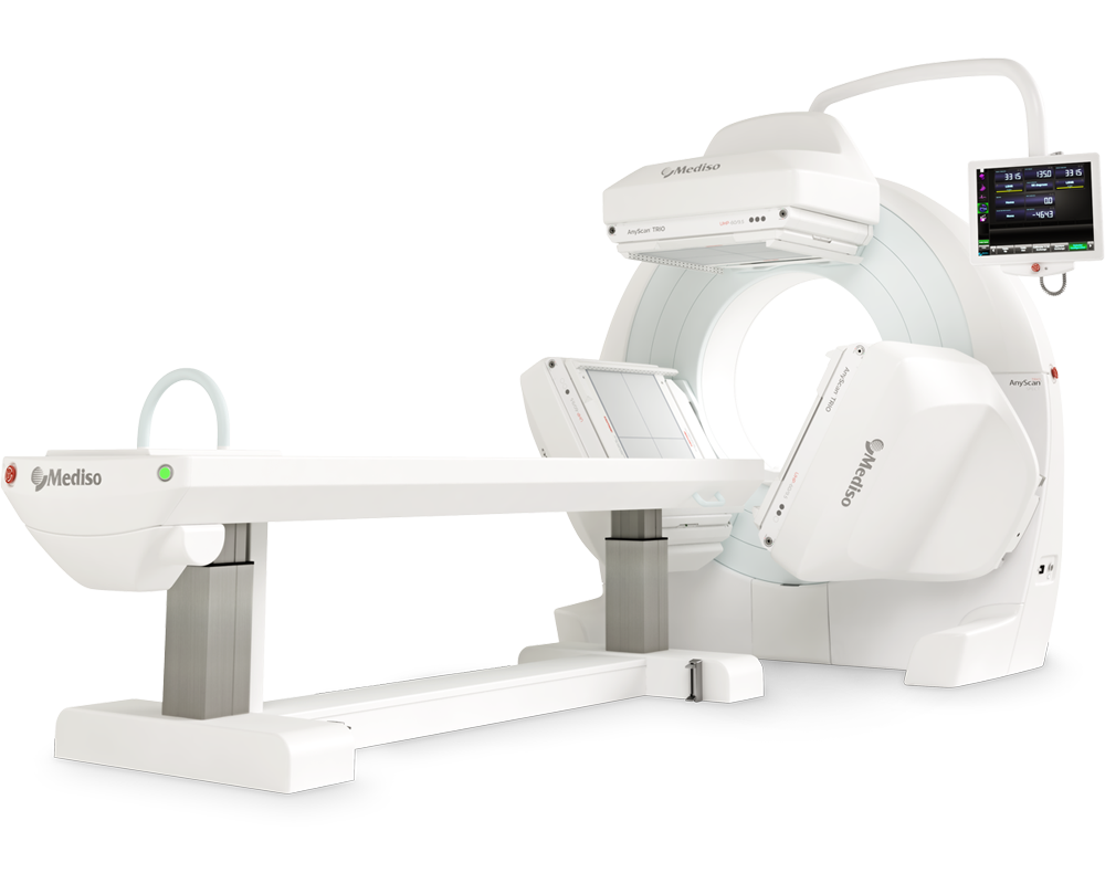
AnyScan® TRIO SPECT
Ultra-fast Triple-NaI-Detector SPECT for wide range of clinical applications
-
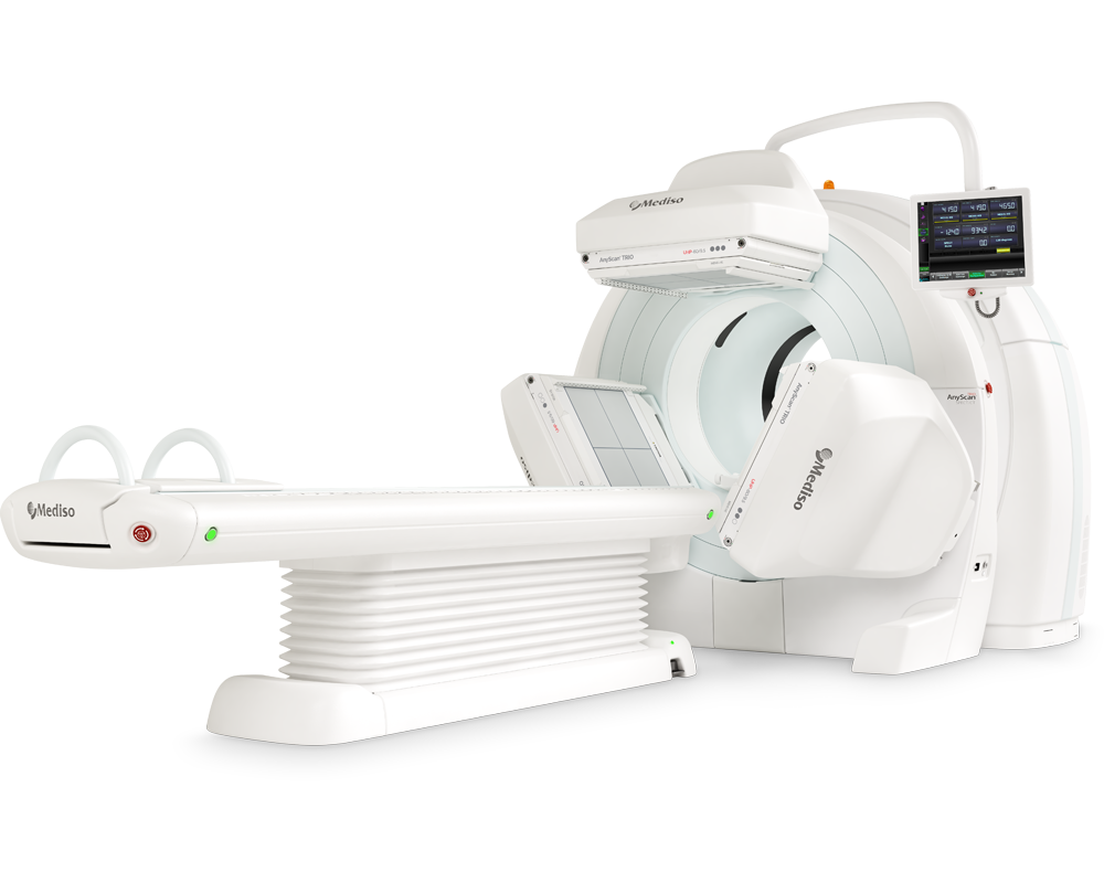
AnyScan® TRIO SPECT/CT
Ultra-fast Triple-NaI-Detector SPECT system for wide range of clinical applicati...
-
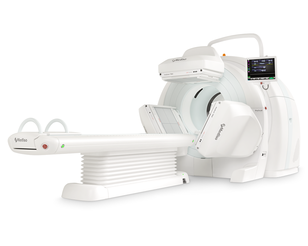
AnyScan® TRIO SPECT/CT/PET
An integrated triple-detector SPECT/CT and PET/CT system in a single-room instal...
-
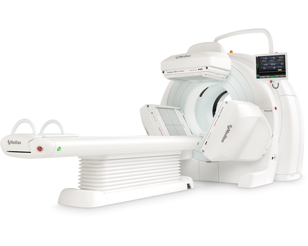
AnyScan® TRIO-TheraMAX SPECT/CT
Jakość Obrazów Teranostycznych i Diagnostycznych na MAXymalnym poziomie


W czym możemy pomóc?
Skontaktuj się z nami aby uzyskać informacje techniczne i / lub wsparcie dotyczące naszych produktów i usług.
Napisz do nas