In Vivo SPECT Imaging of Tc-99m Radiolabeled Exosomes from Human Umbilical-Cord derived Mesenchymal Stem Cells in Small Animals
2024.04.16.
Yi-Hsiu Chung et al, Biomedical Journal, 2024
Summary
Extracellular vesicles derived from human umbilical cord-derived mesenchymal stem cells (UCMSC-EVs) have been postulated to have therapeutic potential for various diseases.
Exosomes are a type of membrane-bound extracellular vesicle (EV) produced in most eukaryotic cells' endosomal compartments. They can contain well-known molecules such as proteins, DNA, RNA, lipids, and metabolites associated with different host cells. Human umbilical cord-derived mesenchymal stem cells (UCMSCs) presenting in the umbilical cord tissue and characterized as self-renewing and multipotent become one of the sources of exosomes. There is currently no clear report on the long-term safety of MSCs, including UCMSCs, in humans despite various clinical trials and results. On the contrary, exosomes derived from UCMSCs (UCMSC-EVs) are thought to possess the characteristics of UCMSCs that can promote tissue regeneration, improve tissue function, regulate the immune system, and exert anti-inflammatory effects. Studies have reported the efficacy of UCMSC-EVs in wound healing and bone regeneration by the local administration, however, their biodistribution and pharmacokinetics are still unclear. UCMSC-EVs have been labelled with dyes, gadolinium, or gold nanoparticles and observed by in vivo imaging, but UCMSC-EVs labelled and quantified with radioisotopes have not yet been studied. For a better understanding of the in vivo properties of UCMSC-EVs, in the present study, these vesicles were first radiolabelled with Technetium-99 m (99mTc-UCMSC-EVs) and evaluated using in vivo single photon emission computed tomography (SPECT) imaging and biodistribution experiments.
The advantages of radioactive UCMSC-EVs include not significantly altering the structure of exosomes, high sensitivity for capturing regions of interest with three-dimensional images as well as elucidating pharmacokinetic information.
SPECT images demonstrated that the liver and spleen tissues mainly took up the 99mTc-UCMSC-EVs. The biodistribution study observed slight uptake in the thyroid and stomach, indicating that 99mTc-UCMSC-EVs was stable at 24 h in vivo. The pharmacokinetic analyses of the blood half-life demonstrated the quick distribution phase (0.85 ± 0.28 min) and elimination phase (25.22 ± 20.76 min) in mice. This study provides a convenient and efficient method for 99mTc-UCMSC-EVs preparation without disturbing their properties. In conclusion, the biodistribution, quick elimination, and suitable stability in vivo of 99mTc-UCMSC-EVs were quantified by the noninvasive imaging and pharmacokinetic analyses, which provides useful information for indication selection, dosage protocol design, and toxicity assessment in future applications.
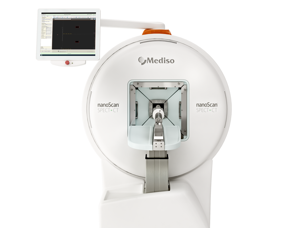
Results from nanoScan® SPECT/CT
Longitudinal in vivo tracking of 99mTc-UCMSC-EVs was carried out using a small animal imaging scanner SPECT/CT (nanoScan, Mediso, Hungary). The SPECT and CT scans were acquired from healthy BALB/c mice after administration of the 99mTc-UCMSC-EVs by intravenous tail injection. 99mTc-NaTcO4 was used as a control.
Images were acquired at 30 min, 1 h, 4 h and 24 h post injection. The 30 min scan time was performed for each experiment except 60 min scan time for 24 h time-point. Multipinhole collimators were used for acquisition of the SPECT images with a 20% energy window and 0.85 mm spatial resolution. The 3D OSEM reconstruction was performed with a 0.26 mm3 voxel size. The SPECT image was subjected to attenuation, scatter correction, and isotope decay correction.
Substantial EV accumulation was observed in the bladder within 30 min post injection, which confirms EV excretion through the urinary tract (bladder activity: 8.71 ± 7.84 %ID/g). Mainly, 99mTc-UCMSC-EVs uptake in the spleen (44.82 ± 9.19%ID/g) and liver (54.65 ± 14.18 %ID/g) occurred after 30 min, and no significant changes in the 99mTc-UCMSC-EVs distribution were observed at further time points, except in the spleen at 24 h (67.57 ± 10.65 %ID/g, p < 0.05). Notably, 99mTc-UCMSC-EVs uptake in the kidneys, lungs, bone, and spine was not observed at 24 h. Significantly high uptake of 99mTcO4- in the thyroid and stomach was observed in a control mouse.

Figure 1. Assessment of the intravenous administration of 99mTc-UCMSC-EVs and comparison with 99mTc-NaTcO4. (a) In vivo SPECT/CT fused representative images of 99mTc-UCMSC-EVs and Na99mTcO4 at various time points post injection. Significant uptake of 99mTc-UCMSC-Evs was found in the liver, spleen, lung, and bone. The elimination route of 99mTc-UCMSC-EVs was shown to be through the urinary tract. Furthermore, there was no observed uptake in the thyroid and stomach for the 99mTc-UCMSC-EVs group compared with the Na99mTcO4 group, indicating that the 99mTc-UCMSC-EVs were stable in vivo. (b) Quantification of 99mTc-UCMSC-EVs in organs/tissues of interest at various time points post injection. The spleen uptake of 99mTc-UCMSC-EVs 24 h post injection was significantly elevated compared with that at previous time points (67.57 ± 10.65%ID/g, *). No 99mTc-UCMSC-EVs were observed in the kidney, lung, bone, spine or bladder.
The half-life of 99mTc-UCMSC-EVs in the blood was determined by measuring radioactivity in serial blood samples at 2 min, 5 min, 10 min, 20 min, 30 min, 1 h, 4 h, and 24 h. The mice were also subjected to SPECT scans to study the pharmacokinetics of 99mTc-UCMSC-EVs. Blood samples were collected from the tail vein of mice under 2% isoflurane anesthesia at several time points post injection. Data were analyzed with a two-phase decay nonlinear regression in GraphPad Prism 6.0. The blood half-life of 99mTc-UCMSC-EVs in the distribution phase and elimination phase was computed. The area under the curve (AUC) of 99mTc-UCMSC-EVs was calculated by radioactivity of blood per ml. The in vivo blood half-life of 99mTc-UCMSC-EVs showed a quick distribution and moderate elimination in the two-compartment model. The blood half-life was 0.85 ± 0.28 min for the distribution phase and 25.22 ± 20.76 min for the elimination phase. The AUC of 99mTc-UCMSC-EVs in blood was 1021.07 ± 397.77 kBq*h/mL from administration to 24 h.
 Figure 2. Pharmacokinetics of 99mTc-UCMSC-EVs (a) The representation of in vivo blood half-life fitting with a two-compartment model. The grey line is the fitting curve. The blood half-life was 0.85 ± 0.28 min in the distribution phase and 25.22 ± 20.76 min in the elimination phase (n = 6). (b) A representative plot of the AUC of 99mTc-UCMSC-EVs is shown from the initial timepoint to 24 h post injection. Collectively, the AUC of 99mTc-UCMSC-EVs in blood was 1021.07 ± 397.77 kBq*h/ml from administration to 24 h (n = 6).
- 99mTc-UCMSC-EVs showed extremely high stability in vitro and in vivo.
- The uptake of 99mTc-UCMSC-EVs after intravenous administration was primarily in the liver and spleen, with minor uptake in the lungs, bone, and spine.
- The 99mTc-UCMSC-EVs circulated in the blood circulation with half-life 25 min and were eliminated through the urinary tract, as demonstrated by SPECT imaging, biodistribution analyses, and pharmacokinetics.
- UCMSC-EVs are expected to be useful in various biomedical applications in the future, thanks to the results of radiolabelling and studying their biodistribution and pharmacokinetics.
Full article on sciencedirect.com
W czym możemy pomóc?
Skontaktuj się z nami aby uzyskać informacje techniczne i / lub wsparcie dotyczące naszych produktów i usług.
Napisz do nas
