Comparison of CT noise reduction performances with deep learning-based, conventional, and combined denoising algorithms
2022.09.22.
Balogh et al., Medical Engineering & Physics, 2022
Abstract
Conventional noise reduction algorithms have been used in image processing for a very long time, but recently, deep learning-based algorithms have been shown to significantly reduce the noise in CT images. In this paper, a comparison of CT noise reduction of a deep learning-based, a conventional, and their combined denoising algorithms is presented. A conventional adaptive 3D bilateral filter and a 2D deep learning-based noise reduction algorithm and a combination of these are compared. For comparison, we used the noise power spectrum and the task transfer function which were measured on original CT images and the effective dose saving factors were also calculated. The noise reduction effect, the noise power spectrum and the task-transfer function are studied using Catphan 600 phantom and 26 clinical cases with more than 100,000 images. We also show that the effect of noise reduction of a 2D deep learning-based algorithm can be further enhanced by using conventional 3D spatial noise reduction algorithms.
Methods
In this paper, we compared an adaptive version of a conventional noise reduction method (bilateral filter) with a deep learning-based convolutional neural network algorithm (DNR; ClariCT.AI, ClariPi) incorporated into a clinical CT (Mediso AnyScan® SC). The degree of noise reduction was also investigated for the combination of the two filters. The performance of DNR has been previously evaluated within several clinical studies.
During the reconstruction process four different kernels were applied; two soft tissue type kernels (Abdomen, Brain) and two high-resolution kernels (Bone, Lung) using the following post-reconstruction protocols of the clinical CT scanner (Mediso AnyScan® TRIO SPECT/CT): AI_Bone, AI_Lung, AI_Abdomen and AI_Brain. We will simply use the following abbreviations for these protocols: Bone, Lung, Abdomen and Brain, respectively. The reconstructed slice thickness was 0.625 mm. The resulting axial slices of FBP reconstruction were also processed with DNR and 3D bilateral filters and their combined one. Throughout this article the unprocessed result of FBP reconstruction is referred to as the original image. Since the deep learning-based algorithm was trained on unprocessed FBP images the output image of the combined filter was the result of the bilateral filter on the output of DNR. Using 0.625 mm thick images helps DNR to have more information in image space, and helps to reduce the blur effect of the 3D bilateral filter. The processed 0.625 mm thickness images were combined in pairs, so that the slice thickness of the final images was 1.25 mm , which is the default post-reconstruction slice thickness of Mediso CT.
The noise reduction effect was studied on 26 clinical cases. All patients were scanned using a clinical CT scanner (AnyScan SPECT/CT, Mediso).
The noise level of the clinical images was measured by an automated algorithm. Measuring the mean noise level in the whole volume characterizes the global noise reduction efficiency more accurately than measuring the noise level in some predefined ROIs in specific areas. By the help of global noise measurement, we measured noise in all reconstructed images using more than 100,000 clinical images during the study.
Results
We investigated 26 clinical cases with different scan parameters. The effect of the bilateral filter, DNR and their combination is demonstrated in the following figures. These figures show reconstructions from very low dose projections.

Reconstructed images of Patient 4 using Abdomen protocol; image size: 512 x 512. Scan parameters were as follows: helical mode; tube voltage, 120 kV; tube current, 10 mA; pitch, 1.5. (A) original image. (B) result of bilateral filtering. (C) Image generated by DNR. (D) Image obtained by the combined noise filtering.

Reconstructed images of Patient 4 using Abdomen protocol; zoomed image size: 256 x 256. Scan parameters were as follows: helical mode; tube voltage, 120 kV; tube current, 10 mA; pitch, 1.5. (A) original image. (B) result of bilateral filtering. (C) Image generated by DNR. (D) Image obtained by the combined noise filtering.

Reconstructed images of Patient 25 using Bone protocol; image size: 512 x 512. Scan parameters were as follows: helical mode; tube voltage, 120 kV; tube current, 70 mA; pitch, 1.5. (A) original image. (B) result of bilateral filtering. (C) Image generated by DNR. (D) Image obtained by the combined noise filtering.

Reconstructed images of Patient 25 using Bone protocol; zoomed image size: 256 x 256. Scan parameters were as follows: helical mode; tube voltage, 120 kV; tube current, 70 mA; pitch, 1.5. (A) original image. (B) result of bilateral filtering. (C) Image generated by DNR. (D) Image obtained by the combined noise filtering.
Conclusion
In this study we measured the noise reduction performances of an adaptive 3D bilateral filter and a deep leaning-based noise reduction algorithm using more than 100,000 images. We have shown that the reciprocal of the noise variance is nonlinear with the mA value of the scan in the case of the images processed by the DNR algorithm, which is an interesting feature of this noise filter. This means that the noise reduction efficiency is better if thinner slices are reconstructed, processed, and averaged for thicker slices after the noise reduction process not just because there is more image information but the DNR is more effective if the image noise is higher, which is true for thinner slice reconstruction. We have also shown that further noise reduction can be achieved by the combination of the conventional and the deep-learning algorithms. The used metrics were TTF, NPS, variance of noise and effective dose saving factor. To calculate the effective dose savings, we assumed that the electronic noise is very low compared to the quantum noise. The data were not specific for any disease or anatomical abnormalities.
Full article on Medical Engineering & Physics
-
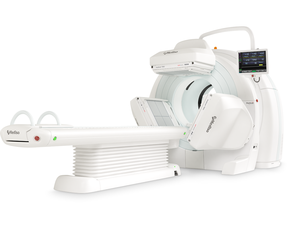
AnyScan® TRIO SPECT/CT
Ultra-fast Triple-NaI-Detector SPECT system for wide range of clinical applicati...
-
AnyScan® TRIO SPECT/CT/PET
An integrated triple-detector SPECT/CT and PET/CT system in a single-room instal...
-
AnyScan® TRIO-TheraMAX SPECT/CT
Jakość Obrazów Teranostycznych i Diagnostycznych na MAXymalnym poziomie


W czym możemy pomóc?
Skontaktuj się z nami aby uzyskać informacje techniczne i / lub wsparcie dotyczące naszych produktów i usług.
Napisz do nas