Functionalization of Radiolabeled Antibodies to Enhance Peripheral Clearance for High Contrast Brain Imaging
2022.10.06.
Eva Schlein et al., 2022, Molecular Pharmaceutics
Summary
The introduction of the positron emission tomography (PET) ligand [11C]Pittsburg compound B ([11C]PiB) about 2 decades ago enabled new possibilities to study and diagnose Alzheimer’s disease (AD), as it binds and visualizes amyloid-beta (Aβ) plaques in the living brain. However, further investigations showed that brain retention of the radiotracer remained static during disease progression and that Aβ pathology in carriers of a specific APP mutation as well as AD patients with predominantly diffuse Aβ plaques cannot be visualized with this method. Furthermore, the soluble forms of Aβ, which are not visualized with [11C]PiB, correlate with neurotoxicity better than Aβ plaques. To image soluble and diffuse aggregates of Aβ, antibody-based imaging techniques could be a suitable approach and an alternative to amyloid radioligands such as [11C]PiB.
Antibody-based imaging techniques have been in focus of research for a while. Despite the high specificity and selectivity, the size of the antibody is often a drawback for its use as an imaging ligand. The majority of antibody ligands are IgG-based and, due to the Fc receptor, the biological half-life of the ligand is long. This long circulation time results in poor contrast, high background signals, and, due to the usage of long-lived radioisotopes, unwanted radiation exposure to normal tissue. To counteract this issue, clearing agents (CAs) can be a useful approach. The task of a CA is to remove radiolabeled antibody from the circulation to enhance the target-to-blood ratio and thereby increase the image quality. Early CAs were based on antibodies raised against the injected antibody ligand, to lower the blood levels of the ligand, or used the avidin–biotin interaction. However, these methods were prone to side-effects, such as immunogenic responses.
An interesting approach to increase antibody clearance, with low side-effects, is the addition of a hexose, such as mannose, to the radioimmunoconjugate. Mannose-modified proteins have been shown to be cleared fast from the circulation by macrophage mannose receptors, which can be found on various tissues, such as endothelial cells in the liver. Bio-orthogonal reactions are a useful approach to efficiently clear antibodies with no known side-effects. A previously developed CA has shown promising results in tumor imaging. This CA is modified with galactose, to actively target the liver via Ashwell receptors on hepatocytes, and functionalized with a tetrazine, which allows in vivo interaction with a trans-cyclooctene (TCO)-functionalized antibody through the inverse-electron demand Diels–Alder (IEDDA) reaction. In this reaction, an electron-deficient diene (tetrazine) and an electron-rich dienophile (TCO) react upon eliminating nitrogen to form a stable bond. This is an irreversible reaction, which is catalyst-free, selective, and mild, as well as compatible with biological systems. Both of the reaction partners, that is, the tetrazine and the TCO, can be introduced into different biomolecules via enzymes or chemical modifications and various applications of the reaction have been explored.
Tha authors have previously developed an antibody, mAb158, which specifically targets Aβ protofibrils, a soluble form of Aβ aggregates. Further development resulted in a bispecific recombinant antibody, RmAb158-scFv8D3, where mAb158 is fused to a single chain variable fragment (scFv) of a rat anti-mouse transferrin receptor (TfR) antibody (8D3), enabling the antibody to cross the blood–brain-barrier up to 80-times more than unmodified antibody. These antibodies have been used for PET or single photon emission computed tomography (SPECT) imaging in transgenic mice with Aβ pathology. Both recombinant, IgG-based antibodies, RmAb158-scFv8D3 and RmAb158, were included in this study.
The aim of this study was to investigate the usage of two different clearing approaches (mannose modification and CA) on the two above-mentioned antibodies to achieve an increase in contrast for brain-imaging purposes.
Results from the nanoScan SPECT/CT
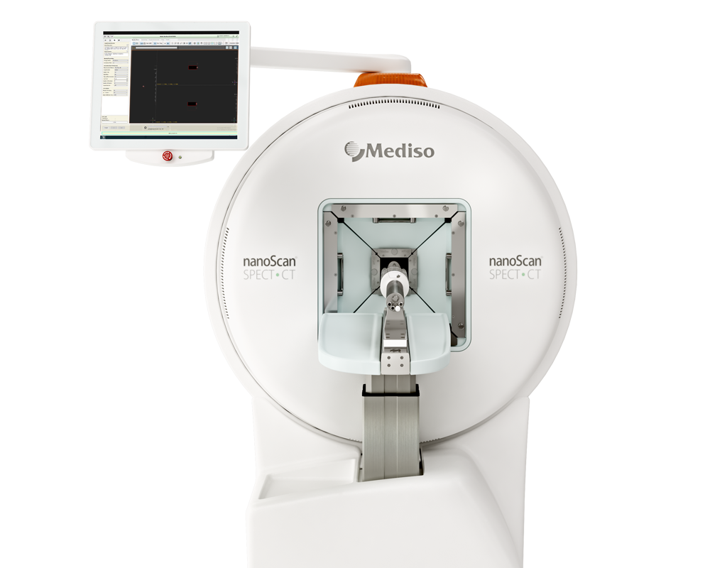
For the SPECT/CT imaging, a subset of mice injected with TCO-[125I]I-RmAb158 were investigated with SPECT imaging, before and after CA administration. SPECT scans were performed 3 or 5 days after injection of radiolabeled antibody, as well as 2 and 24 h after CA administration. Each mouse was scanned maximum three times. Mice were anesthetized with 4% sevoflurane before scanning, positioned on the pre-heated scanner bed of a small animal nanoScan SPECT/CT and scanned for 45–90 min depending on the selected field of view (whole body or head scan). CT was performed with the following settings: 50 kilovoltage peak X-ray, 600 μA, and 480 projections; and CT images were reconstructed using filtered back projections. Iodine-125 γ-emission was collected with an acquisition frame of 2 min. SPECT acquisition data were reconstructed using Nuclide 2.03 software and Tera-TomoTM 3D SPECT reconstructive algorithm with scattering and attenuation correction. SPECT images were reconstructed using 48 iterations into a static image. Reconstructed data were decay-corrected and adjusted for injected dose. Data were visualized in AMIDE v 1.0.4
Figure 6. the impact of the induced clearance on SPECT imaging, TCO-[125I]I-RmAb158 was injected in 18–20 month old tg-ArcSwe and wt mice and scanned at three different time points─before, 2 h after and 24 h after injection of the CA (Figure 6A). As seen in the SPECT images, the signal in the liver increased substantially after CA injection and resulted in an almost complete clearance of the antibody from the body after 24 h (Figure 6AI–III). In comparison, a mouse scanned 5 days after TCO-[125I]I-RmAb158 injection showed a higher overall signal of radioactivity in the body (Figure 6B). Clearance of TCO-[125I]I-RmAb158 from the blood allowed for the visualization of ventricle-associated brain signal from the retained antibody (Figure 6CIII,IV). Sagittal and coronal images of the brain showed a distinct signal in the center of the brain, apparent at 2 h and especially at 24 h after CA administration (Figure 6CII–IV), clearly related to the decreased antibody blood concentration (Figure 6C). This signal likely represents accumulation of the antibody in association with Aβ deposits around the central ventricle, a phenomenon that has previously been observed with SPECT imaging 6–27 days after the injection of [125I]I-RmAb158 in tg-ArcSwe mice. (28) No signal was observed in the brain of TCO-[125I]I-RmAb158 injected wt mice 24 h after CA administration (Figure 6D). The specificity of the SPECT signal was confirmed by ex vivo autoradiography, showing high antibody retention in the tissue of tg-ArcSwe (Figure 6E) but not wt (Figure 6F) mice in the absence of blood.
- This study demonstrates that the principle of induced radioligand clearance, based on the biorthogonal IEDDA reaction, can be used for antibody-based imaging in the CNS.
- With further optimization of antibody design and radiolabeling, this may become a useful strategy to enhance contrast in antibody-based imaging of brain targets.
W czym możemy pomóc?
Skontaktuj się z nami aby uzyskać informacje techniczne i / lub wsparcie dotyczące naszych produktów i usług.
Napisz do nas
