Dose predictions for [177Lu]Lu-DOTA-EGF F(ab′)2 in NRG mice with HNSCC patient-derived tumour xenografts based on [64Cu]Cu-DOTA-EGF F(ab′)2 – implications for a PET theranostic strategy
2021.08.12.
Anthony Ku, Raymond M. Reilly et al., EJNMMI Radiopharmacy and Chemistry
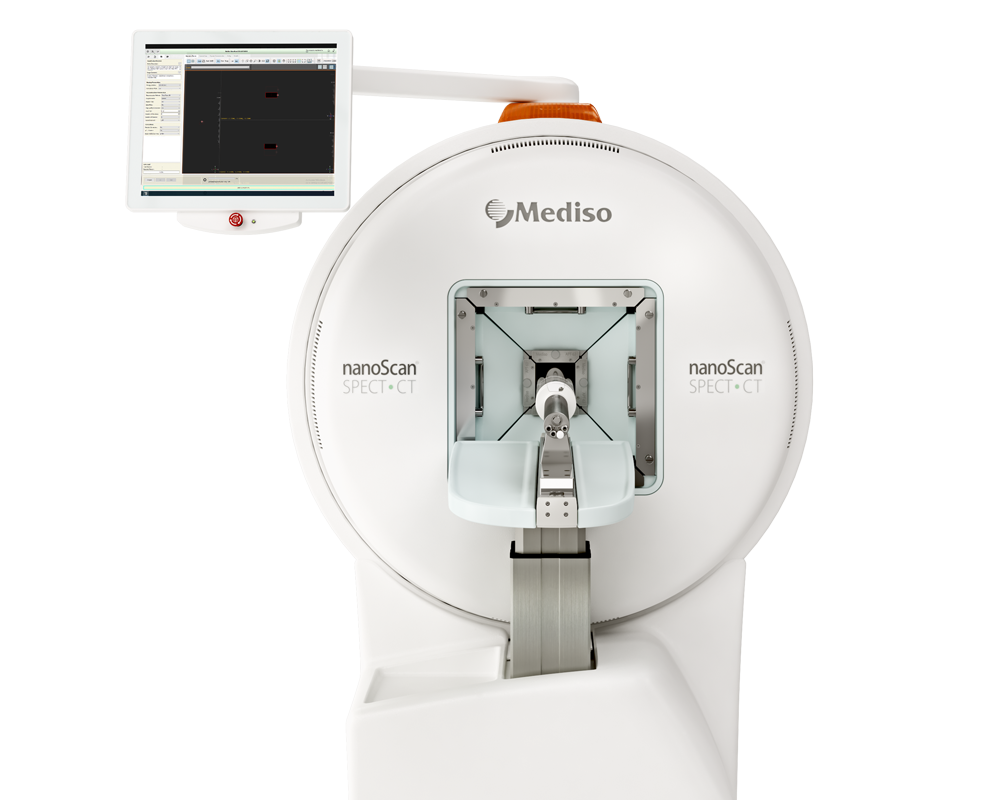
nanoScan® SPECT/CT/PET triple modality system was used to follow a PET and a SPECT radiotracer biodistribution and predict radiation equivalent doses for [177Lu]Lu-DOTA-panitumumab F(ab′)2 radioimmunotherapy.
Background
Epidermal growth factor receptors (EGFR) are overexpressed on many head and neck squamous cell carcinoma (HNSCC). Radioimmunotherapy (RIT) with F(ab')2 of the anti-EGFR monoclonal antibody panitumumab labeled with the βparticle emitter, 177Lu may be a promising treatment for HNSCC. The aim was to assess the feasibility of a theranostic strategy that combines positron emission tomography (PET) with [64Cu]Cu-DOTA-panitumumab F(ab')2 to image HNSCC and predict the radiation equivalent doses to the tumour and normal organs from RIT with [177Lu]Lu-DOTA-panitumumab F(ab')2.
Results from nanoScan® SPECT/CT/PET
- Researchers reported for the first time radiation equivalent dose predictions for [177Lu]Lu-DOTA-panitumumab F(ab´)2 based on biodistribution studies and ROI analysis of microPET/CT images in NRG mice with HNSCC patient-derived xenografts tumours after i.v. administration of [64Cu]Cu-DOTA-panitumumab F(ab´)2.
- Biodistribution (BOD) studies at 6, 24 or 48 h post-injection (p.i.) of [64Cu]Cu-DOTA-panitumumab F(ab')2 (5.5–14.0 MBq; 50 μg) or [177Lu]Lu-DOTA-panitumumab F(ab')2 (6.5 MBq; 50 μg) in NRG mice with s.c. HNSCC patient-derived xenografts (PDX) overall showed no significant differences in tumour uptake but modest differences in normal organ uptake were noted at certain time points.
- [64Cu]Cu-DOTA-panitumumab F(ab')2 and [177Lu]Lu-DOTA-panitumumab F(ab')2 tumour uptake were significantly higher (P < 0.05) than a non anti-EGFR antibody [177Lu]Lu-DOTA-trastuzumab F(ab')2 uptake, demonstrating EGFR-mediated tumour uptake
- Human doses from administration of [177Lu]Lu-DOTA-panitumumab F(ab')2 predicted that a 2 cm diameter HNSCC tumour in a patient would receive 1.1–1.5 mSv/MBq and the whole body dose would be 0.15–0.22 mSv/MBq.

Fig. 3 Posterior whole-body coronal microPET/CT images of a NRG mouse bearing s.c. implanted HNSCC PDX at a. 6 h, b. 24 h or c. 48 h p.i. of [64Cu]Cu-DOTA-panitumumab F(ab´)2. The tumour is shown by the white arrow. At 6 h p.i., the mediastinum (blood pool; white arrowhead), liver (blue arrowhead) and kidneys (broken red circles) are visualized, while at 24 and 48 h p.i., the liver was the only normal organ visualized.

Fig. 4 Posterior whole-body coronal microSPECT/CT images of a NRG mouse bearing s.c. implanted HNSCC PDX at a. 6 h, b. 24 h or c. 48 h p.i. of [177Lu]Lu-DOTA-panitumumab F(ab´)2 or d. 24 h p.i. of irrelevant [177Lu]Lu-DOTA-trastuzumab F(ab´)2. The tumour is shown by the white arrow. At 6 h p.i., the mediastinum (blood pool; white arrowhead), liver (blue arrowhead) and kidneys (broken white circles) were visualized, while at 24 and 48 h p.i., the liver and kidneys were the main normal organs visualized.
Full article published at EJNMMI Radiopharmacy and Chemistry
W czym możemy pomóc?
Skontaktuj się z nami aby uzyskać informacje techniczne i / lub wsparcie dotyczące naszych produktów i usług.
Napisz do nas

