Discovery, optimization and biodistribution of an Affibody molecule for imaging of CD69
2021.09.27.
Jonas Persson, Emmi Puuvuori et al., Scientific Reports, 2021
Summary
The understanding of the immune system has long been an area of intense research. With the emergence of new immunotherapy approaches, it is necessary to find novel non-invasive ways of studying the immune response in different diseases. Similarly, non-invasive tools to evaluate the involvement of the immune system in e.g. COVID-19 is urgently needed for understanding the progression of the disease and assessment of treatment effect. Furthermore, much is still unclear about the autoimmune response in many diseases, including type 1 diabetes (T1D), asthma, rheumatoid arthritis and psoriasis. The field of immunology is in dire need of clinically available biomarker for activated immune cells.
Cluster of differentiation 69 (CD69) is one of the earliest cell surface proteins expressed by activated lymphocytes. It is also part of the lymphocyte proliferation and functions as a signal transmitting receptor. Several other immune cell populations, including monocytes, neutrophils and eosinophils express CD69 in all tissues explored so far. CD69 is constitutively expressed only by mature thymocytes and platelets, and it is not found on the resting circulating leukocytes in humans. In fact, CD69 is also a marker for tissue resident T memory cells due to its ability to suppress tissue egress, by interacting with and internalizing S1P1. Thus, CD69 is a general surface marker for activated immune cells in tissue, with limited presence in the circulation.
Currently, the only available high affinity binders for CD69 are antibodies. Antibodies can be radiolabeled for imaging, but to match the slow clearance, radionuclides with long radioactive half-life and associated radiation dose must be used. Radiation dose can be a limiting factor, especially when imaging vulnerable or young populations. Therefore, an ideal CD69 imaging agent must be significantly smaller than an antibody, while retaining high affinity and specificity. Hence, CD69 may be an interesting target for imaging probes due to the potentially low background binding in blood that otherwise could cause background signal.
Affibody molecules are small (6 kDa) scaffold proteins that consist of three alpha helices with 58 amino acids, which can be randomized to create large libraries from which potential binders may be isolated and further engineered. The benefits of Affibody molecules also include their robustness and due to their small size, they clear out of plasma rapidly and penetrate into tissue. The biodistribution of radiolabeled Affibody molecules will directly influence their potential as in vivo imaging agents. Before embarking on further preclinical evaluation, we performed in vivo imaging in healthy rats to screen the Affibody molecules for lowest background binding in critical tissues (e.g. to optimize image contrast) as well as kidney retention (e.g. to choose the variant with optimal radiation dosimetry).
The aim of this study was to generate, characterize and optimize an Affibody molecule targeting CD69 for further development as an imaging agent of activated immune cells.
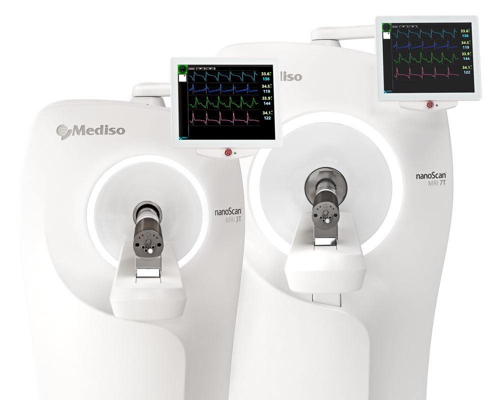
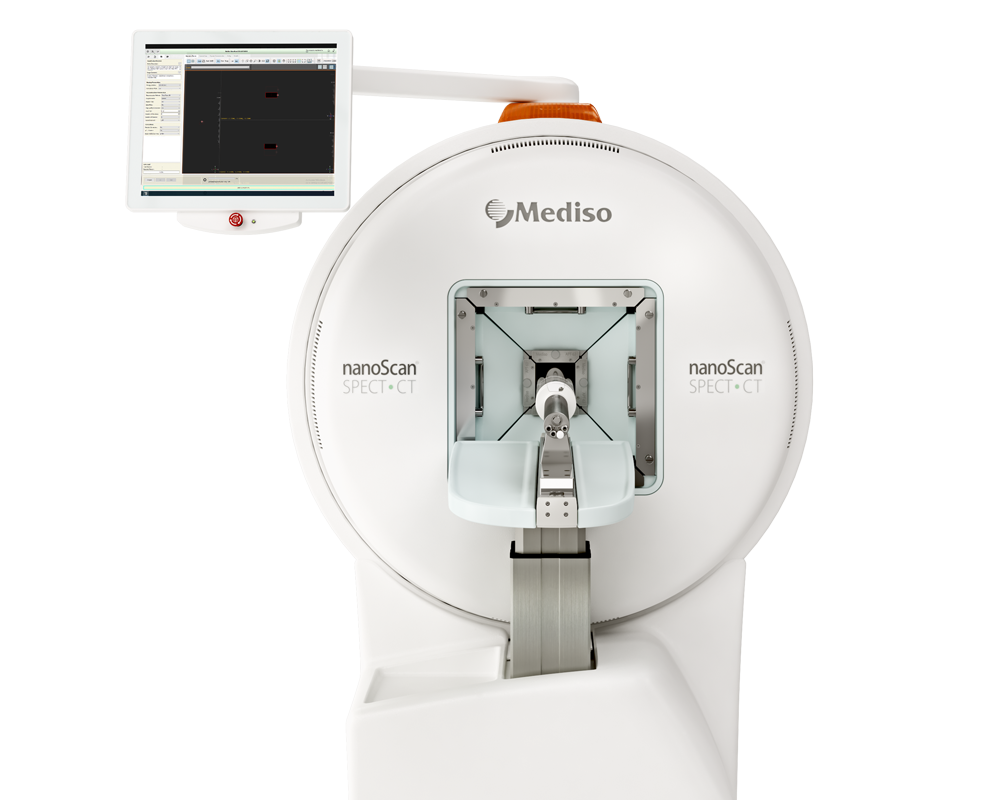
Results from the nanoScan SPECT/CT and nanoScan PET/MRI 3T
For the imaging studies, 111In-DOTA-ZCD69:# (affinity matured) variant biodistribution in healthy rats was assessed by Single-photon emission computed tomography (SPECT-CT) imaging. Sprague–Dawley rats (n = 15 in total, n = 3 per Z variant, male, weight 318 ± 43 g) were injected in the lateral tail-vein with approximately 8 MBq of indium-111 labeled Z variant (111In-DOTA-ZCD69:2: 3.9 ± 1.6 MBq; 111In-DOTA-ZCD69:4: 12.6 ± 4.6 MBq; 111In-DOTA-ZCD69:6: 5.5 ± 1.3 MBq; 111In-DOTA-ZCD69:8: 8.0 ± 2.6 MBq; 111In-DOTA-ZCD69:12: 9.9 ± 2.6 MBq, 111In-DOTA-ZTAQ: 2.7 ± 1.4 MBq).
The examination by SPECT/CT was carried out immediately post injection (0 h) as well as 3 h, 20 h, 48 h and 72 h post injection. For each scan, the animal was anesthetized and positioned by a whole-body CT acquisition. Next, a 20-min whole body static SPECT examination was performed. Some the animals were then moved to the nanoScan PET/MRI scanner via the detachable bed, and examined by MRI. During each imaging session, the rat was sedated by gas anesthesia (sevoflurane 5% initially and afterwards 3% to maintain anesthesia) through a facemask. Temperature was maintained by warm air supply integrated in the scanner bed.
CT acquisition was performed before all SPECT examinations for anatomical co-registration (semicircular multi-field-of-view; duration per bed 7:46 min; 3 rotations; scan length 231.66 mm; 480 projections; binning 1:4; 50 kV; 600 µA; voxel size 0.25 × 0.25 × 0.25 mm). SPECT were acquired using the energy windows suitable for indium-111 emission (energy map: primary peak 245.35 keV, secondary peak 171.30 keV), and reconstructed using an iterative algorithm (iterations/subsets 48/3).
SPECT image analysis for all time-points was performed using the Nucline software (Mediso, Hungary). The kidneys, liver, heart, lung and muscle were segmented directly on SPECT images using co-registered CT projections as support. The uptake values were decay corrected to the time of administration and converted to standardized uptake values (SUV) by correcting for animal weight and administered amount of 111In-DOTA-ZCD69:# variant.
Fig. 5. shows representative coronal SPECT, CT and MRI images showing biodistribution and lymph node (red arrows) uptake 24 h post administration of 111In-DOTA- ZCD69:6 (A) and negative control 111In-DOTA- ZTAQ (B). After injection in rats, all indium-111 labeled Z variants exhibited rapid excretion though the kidneys and washout from most tissues (Fig. 5A–E, Fig. 6, Supplementary Figs. 13–17). The uptake and retention in the kidney cortex tended to differ between the Z variants, with 111In-DOTA-ZCD69:2 demonstrating the highest kidney uptake, followed by 111In-DOTA-ZCD69:6 (Fig. 5C). 111In-DOTA-ZCD69:4 and 111In-DOTA-ZCD69:8 had intermediate kidney uptake, while 111In-DOTA-ZCD69:12 exhibited the lowest uptake at the 48 h time point (Fig. 5C). For the background binding in liver and muscle tissue, 111In-DOTA-ZCD69:4 demonstrated the lowest binding, followed by 111In-DOTA-ZCD69:8 (Fig. 5D,E). 111In-DOTA-ZCD69:2, 111In-DOTA-ZCD69:6 and 111In-DOTA-ZCD69:12 all had higher background binding in liver and muscle (Fig. 5D,E). The control, non-binding affibody 111In-DOTA-ZTAQ demonstrated a similar biodistribution pattern including renal excretion (Fig. 5B).
- In summary, variant ZCD69:4 displayed the optimal properties, both regarding stability, affinity and biodistribution, and was selected as lead compound for further development of a novel class of CD69 imaging agents.
- Future studies will focus on functionalization of ZCD69:4 to allow radiolabeling with positron emitting nuclides (e.g. 18F and 68 Ga) to generate a construct useful for in vivo PET imaging of CD69.
W czym możemy pomóc?
Skontaktuj się z nami aby uzyskać informacje techniczne i / lub wsparcie dotyczące naszych produktów i usług.
Napisz do nas

