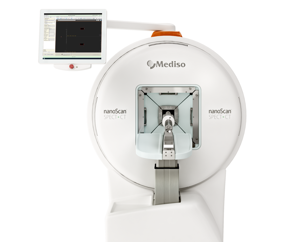
Degree of dopaminergic degeneration measured by 99mTc-TRODAT-1 SPECT/CT imaging
2018.07.13.
Ling Lin et al, Neural Regeneration Reaserch, 2018
Summary
To prevent and treat Parkinson’s disease (PD) in its early stages, it is essential to detect the degree of early dopaminergic neuron degeneration. Dopamine transporters (DAT) in the striatum regulate synaptic dopamine levels, and affect locomotor activity in PD. In addition, dopaminergic neuron degeneration in the substantia nigra pars compacta (SNc) results in a decrease in the density of DAT in the striatum. DAT binding can be used to assess dopamine (DA) function and SPECT scans using 99mTc-TRODAT-1 (a 99mTc-labeled tropane derivative) can be used to image DAT, therefore, a measurement of DAT density in the DA nerve terminal can indicate the severity of DA neuronal loss. Although the decrease in striatal DAT density has been described in clinical PD patients and 99mTc-TRODAT-1 SPECT imaging has been demonstrated as a useful method for diagnosing PD in its early stages, only few reports have been published regarding the association between the degree of 6-hydroxydopamine (6-OHDA)-induced DAergic degeneration and in vivo 99mTc-TRODAT-1 SPECT/CT imaging.
In the present study, DAT imaging was used with 99mTc-TRODAT-1 SPECT/CT to evaluate DAT density in the striatum of unilaterally 6-OHDA-lesioned rats. To mimic the progressive neuronal loss in PD patients, different degrees of nigrostriatal DA depletion were generated by injecting different doses of 6-OHDA. The severity of the lesions was tested by monitoring animal motor behavior and using tyrosine hydroxylase (TH) immunohistochemistry.
The results showed that striatal 99mTc-TRODAT-1 binding was significantly diminished both in the ipsilateral and the contralateral sides in the 4 and 8 μg 6-OHDA groups. There were significant correlations between DAT 99mTc-TRODAT-1 binding in the ipsilateral striatum and the amount of tyrosine hydroxylase immunoreactive neurons in the ipsilateral substantia nigra in the 2, 4, and 8 μg 6-OHDA groups at 8 weeks post-lesion (r = 0.899, P < 0.01). These findings indicate that striatal DAT imaging using 99mTc-TRODAT-1 is a useful technique for evaluating the severity of dopaminergic degeneration.
Results from nanoScan SPECT/CT
Lesioning DA neurons was performed by stereotaxic infusion of 6-OHDA (6-OHDA hydrochloride; 2, 4, or 8 μg) to the right medial forebrain bundle of 32 male adult Sprague-Dawley rats.
Eight weeks after 6-OHDA injection, rats were injected with 99mTc-TRODAT-1 (148–185 MBq/300 μL) via the tail vein. The changes in DAT density in the striatum were detected by a nanoScan SPECT/CT: CT data were acquired using an X-ray voltage biased to 50 kVp with a 670 μA anode current, and the projections were 720°. At 30 minutes post injection, SPECT images were acquired with low-energy, high-resolution collimators. Emission data were acquired in a 256 × 256 matrix size through 360° rotation at 15° intervals for 30 seconds per angle step.
For the analysis of striatal 99mTc-TRODAT-1 binding, the reconstructed image with the highest signal in the striatum was summed together with its two adjacent slices as a single composite image. Regions of interest were drawn in the striatum in each hemisphere. The DAT radioactivity [99mTc-TRODAT-1 binding (bq/mm3)] was counted and corrected for background activity from the cerebellum.
After SPECT imaging, the degree of DAergic neuron loss in the SNc was confirmed by immunohistochemistry.
Results show
- Unilateral injection of 6-OHDA resulted in a significant decrease in striatal 99mTc-TRODAT-1 binding (bq/mm3), both in the contralateral and the ipsilateral sides, in the 4 and 8 μg 6-OHDA groups compared with the corresponding side in the control or vehicle group at 8 weeks post-lesion (C and D)
- When lesions were induced by 2 μg 6-OHDA, the amount of 99mTc-TRODAT-1 binding on the ipsilateral side was significantly reduced compared with the same side in the control group (B)


- Degree of DAergic neuron loss in the SNc was confirmed by immunohistochemistry: at 8 weeks post-lesion, the remaining numbers of TH-immunoreactive neurons in the ipsilateral SNc were significantly lower than those on the contralateral side. There was a positive correlation between DAT radioactivity in the ipsilateral striatum and the numbers of TH-immunoreactive neurons in the ipsilateral SNc at 8 weeks post-lesion. The Pearson’s correlation coefficient was 0.899 at the 0.01 level


W czym możemy pomóc?
Skontaktuj się z nami aby uzyskać informacje techniczne i / lub wsparcie dotyczące naszych produktów i usług.
Napisz do nas