An indium-111-labelled membrane-targeted peptide for cell tracking with radionuclide imaging
2022.10.19.
Johanna Pruller et al, RSC Chemical Biology, 2023
Summary
Cell labelling agents that enable longitudinal in vivo tracking of administered cells will support the clinical development of cell-based therapies. Radionuclide imaging with gamma and positron-emitting radioisotopes can provide quantitative and longitudinal mapping of cells in vivo. To make this widely accessible and adaptable to a range of cell types, new, versatile and simple methods for directly radiolabelling cells are required. We have developed [111In]In-DTPA-CTP, the first example of a radiolabelled peptide that binds to the extracellular membrane of cells, for tracking cell distribution in vivo using Single Photon Emission Computed Tomography (SPECT). [111In]In-DTPA-CTP consists of (i) myristoyl groups for insertion into the phospholipid bilayer, (ii) positively charged lysine residues for electrostatic association with negatively charged phospholipid groups at the cell surface and (iii) a diethylenetriamine pentaacetate derivative that coordinates the g-emitting radiometal, [111In]In3+. [111In]In-DTPA-CTP binds to 5T33 murine myeloma cells, enabling qualitative SPECT tracking of myeloma cells’ accumulation in lungs immediately after intravenous administration. This is the first report of a radiolabelled cell-membrane binding peptide for use in cell tracking.
Results from nanoScan® SPECT/CT
SPECT imaging with [111In]In-DTPA-CTP
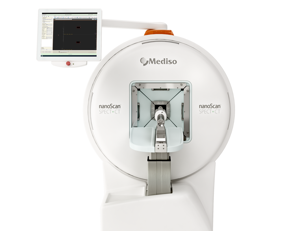
Previous studies have demonstrated that in mice, 5T33 murine myeloma cells localise in the lungs 0–2 h post-injection (PI), followed by migration to the liver, spleen and bone marrow within 24 h. To probe the ability of [111In]In-DTPA-CTP to image the distribution of administered cells in vivo, 5T33 cells labelled with [111In]In-DTPA-CTP (4x10⁶ cells, 7–10 MBq indium-111) were administered intravenously via the tail vein to immunodeficient NSG male mice. A SPECT/CT scan was acquired for 2 h immediately after injection using nanoScan SPECT/CT system (Mediso Medical Imaging Systems) (Figure 1a). For comparative purposes, (i) cell-free [111In]In-DTPA-CTP (2.5 µg peptide, 3–3.3 MBq indium-111) (Figure 1b) and (ii) 5T33 cells labelled with [111In]In-(oxine)3 (4 x 10⁶ cells, 1.8–2.4 MBq indium-111) (Fig. 1c) were administered to mice in parallel, with a 2 h SPECT/CT scan acquired immediately post-injection. The whole-body SPECT scan time was 30 min x 4, (conducted sequentially) with a frame time of 40s (using a 4-head scanner with 4 x 9 (1.4mm) pinhole collimators in helical scanning mode) followed by a helical CT (45kVP X-ray source, 1000 ms exposure time in 180 projections over 7.5 min). After this, the animals were allowed to recover. SPECT/CT scans were acquired again, 1 day PI, after which all animals were culled and organs/tissues harvested, weighed and radioactivity counted using a gamma counter. SPECT/CT images were reconstructed in a 256 x 256 matrix using HiSPECT (ScivisGmbH) and visualised and quantified using VivoQuant v.3.0 software (InVicro LLC.).
The results show that:
- [111In]In-(oxine)3-labelled 5T33 cells localised to the lungs and then the liver 0–2 h PI, consistent with prior studies (Figure 1c).
- Mice injected with cell-free [111In]In-DTPA-CTP exhibited indium-111 activity in the blood pool and liver 0–2 h PI (Figure 1b).
- Mice injected with [111In]In-DTPA-CTP-labelled cells exhibited indium-111 activity in the lungs, liver and blood pool (Figure 1a).
- For the latter group of mice, SPECT/CT data indicated that although some indium-111 radioactivity accumulated in the lungs 0–30 min PI, this decreased over the course of the next 90 min.
- In contrast to mice administered [111In]In-(oxine)3-labelled 5T33 cells, there was significantly greater indium-111 localisation in the liver relative to the lungs.
- At 1 day PI, SPECT/CT scans were acquired and all animals were culled, with organs harvested for ex vivo radioactivity counting (Figure 2).
- There were high concentrations of indium111 in the liver and spleen of mice administered [111In]In- (oxine)3-labelled 5T33 cells or [111In]In-DTPA-CTP-labelled cells, with no statistically significant differences in these organs across these two groups.
It is suggested that some of the initial lung indium-111 uptake observed for animals administered [111In]In-DTPA-CTP-labelled cells is a result of accumulation of [111In]In-DTPA-CTP-labelled 5T33 cells to the lungs. However, over the course of 0–2 h PI, [111In]In-DTPA-CTP dissociates from 5T33 cells, resulting in indium-111 activity in the blood pool and liver 0–2 h PI, similar to the case of mice administered cell-free [111In]In-DTPA-CTP. In contrast, [111In]In-(oxine)3-labelled cells migrate from the lungs to the liver and spleen over a longer time frame, with lower blood pool and liver localisation over 0–2 h PI (Figure 1) and high liver and spleen localisation by 1 day PI (Figure 2). This is consistent with data from prior studies. Although animals administered [111In]In-DTPA-CTP-labelled 5T33 cells demonstrate high indium-111 radioactivity accumulation in the liver and spleen 1 day PI, this is likely to be a result of accumulation of [111In]In-DTPA-CTP that has dissociated from 5T33 cells.

Figure 1. SPECT/CT maximum intensity projections and 111In concentrations in specific organs for mice administered (a) [111In]In-DTPA-CTP-labelled 5T33 cells (SPECT scale of 0.8–8%ID/g), (b) [111In]In-DTPA-CTP (cell-free) (SPECT scale of 1.6–8%ID/g) and (c) [111In]In-(oxine)3-labelled 5T33 cells (SPECT scale of 1.6–16%ID/g), acquired 0–30 min, 30–60 min, 60–90 min and 90–120 min post-injection.

Figure 2. SPECT/CT maximum intensity projections of mice, acquired 1 day post-injection of (a) [111In]In-DTPA-CTP-labelled 5T33 cells; (b) [111In]InDTPA-CTP (cell-free); (c) [111In]In-(oxine)3-labelled 5T33 cells (scale of 0.6–6%ID/g for all images). In all images, indium-111 is largely concentrated in the liver and spleen. (d) Ex vivo biodistribution of 111In radioactivity in animals administered [111In]In-radiotracers. Error bars represent standard deviation. For animals administered [111In]In-DTPA-CTP-labelled 5T33 cells and [111In]In-(oxine)3-labelled 5T33 cells, n = 3, for animals administered cell-free [111In]In-DTPA-CTP, n = 2.
This first cell tracking study on a radiolabelled cell membrane binding peptide has shown that [111In]In-DTPA-CTP-labelled myeloma cells can be qualitatively tracked to the lungs using SPECT imaging immediately after intravenous administration. Although [111In]In-DTPA-CTP dissociates from cell membranes within 1–2 h post-administration of cells and thus falls short of cell adhesion stability requirements for quantitative long-term cell tracking with SPECT, this proof-of-principle study demonstrates the simplicity and feasibility of using synthetic cell membrane binding/penetrating peptides for radiolabelling cells.
Full article on pubs.rsc.org
W czym możemy pomóc?
Skontaktuj się z nami aby uzyskać informacje techniczne i / lub wsparcie dotyczące naszych produktów i usług.
Napisz do nas
