Comparison of tumor‑targeting properties of directly and indirectly radioiodinated designed ankyrin repeat protein (DARPin) G3 variants for molecular imaging of HER2
2019.01.25.
Anzhelika Vorobyeva et al, International Journal of Oncology, 2019
Summary
Evaluation of human epidermal growth factor receptor 2 (HER2) expression levels in breast and gastroesophageal cancer is used for the stratification of patients for HER2‑targeting therapies. The use of radionuclide molecular imaging may facilitate such evaluation in a non‑invasive way. Designed ankyrin repeat proteins (DARPins) are engineered scaffold proteins with high potential as probes for radionuclide molecular imaging. DARPin G3 binds with high affinity to HER2 and may be used to visualize this important therapeutic target. Studies on other engineered scaffold proteins have demonstrated that selection of the optimal labeling approach improves the sensitivity and specificity of radionuclide imaging. The present study compared two methods of labeling G3, direct and indirect radioiodination, to select an approach providing the best imaging contrast. G3‑H6 was labeled with iodine‑124, iodine‑125 and iodine‑131 using cysteine, G3‑GGGC, was site‑specifically labeled using [125I]I‑iodo‑[(4‑hydroxyphenyl)ethyl]maleimide (HPEM). The two radiolabeled G3 variants preserved binding specificity and high affinity to HER2‑expressing cells. The specificity of tumor targeting in vivo was demonstrated. Biodistribution comparison of [131I]I‑G3‑H6 and [125I]I‑HPEM‑G3‑GGGC in mice, bearing HER2‑expressing SKOV3 xenografts, demonstrated an appreciable contribution of hepatobiliary excretion to the clearance of [125I]I‑HPEM‑G3‑GGGC and a decreased tumor uptake compared to [131I]I‑G3‑H6. The direct label provided higher tumor‑to‑blood and tumor‑to‑organ ratios compared with the indirect label at 4 h post‑injection. The feasibility of high contrast PET/CT imaging of HER2 expression in SKOV3 xenografts in mice using [124I]I‑G3‑H6 was demonstrated. In conclusion, direct radioiodination is the preferable approach for labeling DARPin G3 with iodine‑123 and iodine‑124 for clinical single photon emission computed tomography and positron emission tomography imaging.
Results from nanoScan SPECT/CT and nanoScan PET/MRI
Female BALB/c nu/nu mice had an adaptation period of 1 week prior to the start of the experimental procedures. For the implantation of tumors, 107 of SKOV3 cells with high HER2 expression or 5x10⁶ of A431 cells with low HER2 expression in 100 μl media were subcutaneously injected in the right hind leg. The experiments were performed two and a half weeks after implantation.
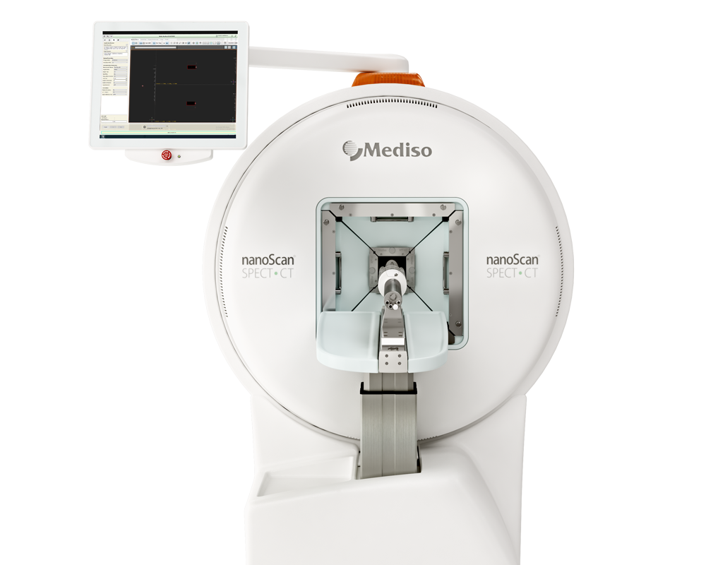
The SPECT study was performed using 125I‑labeled G3‑H6. A mouse bearing SKOV3 xenografts with high HER2 expression was injected with [125I]I‑G3‑H6 (19.7 MBq). The SPECT imaging was performed using nanoScan SPECT/CT at 1, 2 and 4 h post‑injection. The acquisition time was 15 min. CT scans were acquired using the following parameters: X‑ray energy peak of 50 keV; 670 μA; 480 projections; and 5.26 min acquisition time. A mouse bearing A431 xenografts with low HER2 expression was injected with [125I]I‑G3‑H6 (19.7 MBq). The imaging was performed at 4 h post‑injection using the same settings as those for the mouse bearing the SKOV‑3 xenograft. SPECT raw data were reconstructed using Tera‑Tomo™ 3D SPECT reconstruction technology (version 3.00.020.000; Mediso Medical Imaging Systems Ltd.): Normal dynamic range; 48 iterations; 1 subset.
Whole‑body PET imaging was performed using nanoScan PET/MRI. A mouse bearing SKOV3 xenografts was injected with [124I]I‑G3‑H6 (4.7 MBq) and imaged at 4 h pi. The PET scan was performed for 90 min; subsequently, the CT scan was performed using nanoScan SPECT/CT using the same bed position as for the PET scan. The CT scan was acquired using the same parameters as for the SPECT/CT images and the CT data were reconstructed in the same way. PET data were reconstructed using the Tera‑Tomo 3D reconstruction engine. PET and CT files were fused using Nucline 2.03 Software and are presented as maximum intensity projections (MIP) in the RGB color scale.
- The SPECT/CT data revealed a high activity accumulation in SKOV‑3 xenografts with already‑high HER2 expression at 1 h following injection (Fig 7A)
- Appreciable activity accumulation was visualized in the kidneys and stomach (Fig 7A)
- 2 h after injection (Fig 7B) and 4 h after injection (Fig 7C), the high activity uptake in the tumor remained, but the activity in the kidneys was considerably reduced

Figure 7. Imaging of HER2 expression in mice bearing SKOV3 xenografts at 1, 2 and 4 h. Micro‑single photon emission computed tomography/computed tomography imaging of human epidermal growth factor receptor 2 expression in BALB/C nu/nu mice bearing SKOV3 xenograft at (A) 1, (B) 2 and (C) 4 h post‑injection using [125I]I‑G3‑H6. The uptake of radiocatabolites by Na/I‑symporters was blocked by addition of KI in drinking water
- The uptake in the A431 xenografts with low HER2 expression (Fig 8B) was lower compared with that in the SKOV3 xenograft, as expected (Fig 8A), while the uptake in other tissues followed the same pattern as in the mouse with the SKOV3 xenograft

Figure 8. Comparative imaging of high (SKOV3) and low (A431) HER2-expressing xenografts in mice. Micro‑single photon emission computed tomography/computed tomography imaging of human epidermal growth factor receptor 2 expression in BALB/C nu/nu mice bearing (A) SKOV3 and (B) A431 xenografts at 4 h post‑injection using [125I]I‑G3‑H6. The uptake of radiocatabolites by Na/I‑symporters was blocked by addition of KI in drinking water. S, stomach; K, kidneys; T, tumor
- PET/CT imaging with [124I]I‑G3‑H6, clearly visualized the HER2‑expressing SKOV‑3 xenograft at 4 h pi (Fig 9). Low accumulation of activity was observed in other organs, with the exception of the stomach contents in the upper abdomen

Figure 9. MicroPET/computed tomography imaging of human epidermal growth factor receptor 2 expression in BALB/C nu/nu mouse bearing SKOV3 xenograft at 4 h post‑injection using [124I]I‑G3‑H6. The uptake of radiocatabolites by Na/I‑symporters was blocked by addition of KI in drinking water. The area corresponding to the activity in urinary bladder was removed from the PET image during reconstruction to enable better visualization of tumor uptake. S, stomach; T, tumor; PET, positron emission tomography
Full article on spandidos-publications.com
Hogyan segíthetünk Önnek?
További termékinformációkért, vagy támogatásért keresse szakértőinket!
Vegye fel a kapcsolatot

