Inhibition of HIF-1α by Atorvastatin During 131I-RTX Therapy in Burkitt’s Lymphoma Model
2020.05.11.
Eun-Ho Kim et al., MDPI Cancers, 2020
Summary
Radioimmunotherapy (RIT) using radiolabeled monoclonal antibodies served as a targeted therapy for the treatment of relapsed and refractory non-Hodgkin lymphomas (NHL). CD20 is an extracellular surface protein expressed in most human B-cell-lineage malignancies; anti-CD20 antibodies are known for their potential ability to target NHL. Rituximab (RTX) is a monoclonal antibody for the CD20 antigen. RIT using 131I-RTX has already been reported for relapsed or refractory indolent patients with NHL. In RIT of NHL, the targeting properties of anti-CD20 antibodies are explored via conjugation of the antibodies with radioactive isotopes. Despite the success of RIT, certain patients treated with conventional RIT still relapse.
Although HIF-1α is an important biomarker during radiation therapy in solid tumors, its role in NHL is unclear. Notably, it has been reported that overall survival of HIF-1α-positive patients was superior to that of HIF-1α-negative patients with diffuse large B cell lymphoma. To the best of our knowledge, the role of HIF-1α in patients with Burkitt's lymphoma has not been reported to date.
Atorvastatin (ATV) is used to lower cholesterol in the treatment of hypercholesterolemia. The authors have previously demonstrated that ATV enhanced the anticancer effect when used in combination therapy with trastuzumab.
In this study, the authors have investigated whether ATV downregulated tumor radio-resistance and enhanced the anticancer effect of 131I-RTX in BL Raji xenograft mouse models.
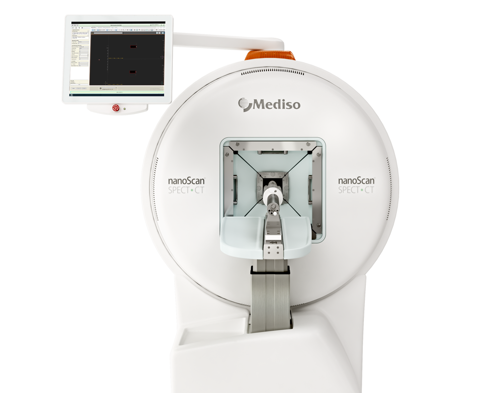
Results from the nanoScan SPECT/CT
For the small animal imaging, the authors have used a nanoScan SPECT/CT, to follow the uptake of 131I-RTX in the Raji cell tumors, and differentiate the effect of using ATV prior to the acquisitions.
When the tumor size reached ~ 200 mm3, ATV (12 μg/day in PBS) was orally administered daily for a total of 10 days; PBS was administered to the control group. 131I-RTX (150 μg, 12.1–14.6 MBq/200 μL) was intravenously injected after 5 days of administration of ATV or PBS. SPECT data were obtained at 2, 24, 48, and 72 h after the injection of 131I-RTX.
Figure 2. shows the main results from the in vivo studies. (A) Representative SPECT/CT images of Raji-xenografted mice were acquired at 2, 24, 48, and 72 h after injection of 131I-RTX (upper row) and ATV plus 131I-RTX (lower row). White dotted circles indicate the tumor region. (B) The quantification of 131I-RTX accumulation in tumors is represented by the tumor to blood ratio at each time point (*p < 0.05). The data are the mean ± SD from five independent mice. (C) Autoradiography of 131I-RTX in Raji tumors was conducted after the acquisition of SPECT images (upper row). (D) The total accumulation of 131I-RTX per tumor tissue (**p < 0.005). The data are the mean ± SD from ten independent images.
- Immuno-SPECT images demonstrated a higher uptake of 131I-RTX in tumors of the ATV-treated group than that of the PBS group (Figure 2a). The tumor to blood ratios (%) of 131I-RTX in ATV group were 1.4-fold and 1.2-fold higher than those in the PBS group at 48 and 72 h, respectively, after injection (Figure 2b; *p < 0.05).
- The findings of the authors have suggested that ATV + 131I-RTX therapeutic regimen is a promising strategy for enhancing the potency of 131I-RTX therapy in poorly responding patients and those with radio-resistance.

Related products
Hogyan segíthetünk Önnek?
További termékinformációkért, vagy támogatásért keresse szakértőinket!
Vegye fel a kapcsolatot
