Bispecific GRPR-Antagonistic Anti-PSMA/GRPR Heterodimer for PET and SPECT Diagnostic Imaging of Prostate Cancer
2019.09.14.
Bogdan Mitran et al., MDPI Cancers, 2019
Summary
Until recently, the diagnosis of prostate cancer (PCa) was based on measuring the concentration of prostate-specific antigen (PSA) in blood, a pathological examination of biopsy material, positron emission tomography (PET) imaging of metabolic activity using 18F-flourodeoxyglucose (FDG) or 11C-acetate, and proliferation activity using radiolabeled choline. Bombesin agonistic analogues for the imaging of gastrin-releasing peptide receptor (GRPR) expression were also proposed, but their clinical implementation was limited due to their strong physiological action.
The development of small-molecular-weight prostate-specific membrane antigen (PSMA)-targeting derivatives of urea was a big breakthrough in the diagnosis of PCa, however despite the apparent success, the imaging of PCa lesions requires further improvement because of the sensitivity of the imaging.
Another relevant target for the diagnostic imaging of PCa is GRPR, which is expressed in earlier stages of PCa. The gallium-68-labeled GRPR antagonist RM2 was used in several clinical studies, with promising results.
In this study, Mitran et al. report the synthesis of a new PSMA/GRPR-targeting heterodimer that includes a Glu-Ureido PSMA-binding moiety and GRPR antagonist RM26 coupled via a glutamic acid bearing NOTA chelator. The aim of this study was to preclinically evaluate the gallium- and indium-labeled heterodimer in terms of the binding properties to PSMA and GRPR, cellular processing, in vivo targeting, and biodistribution, as well as the imaging properties.
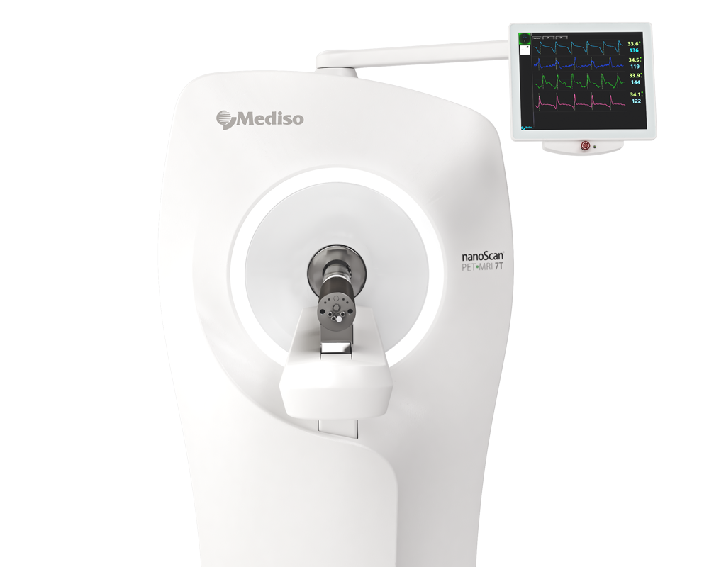
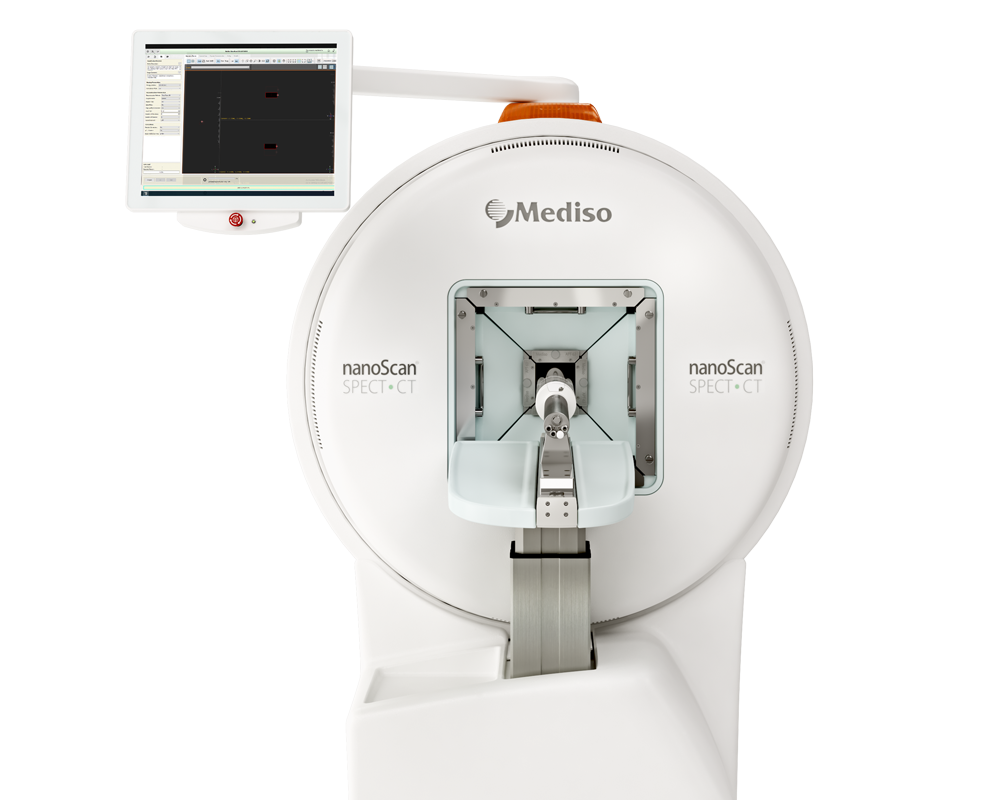
Results from the nanoScan® PET/MRI 3T and nanoScan® SPECT/CT
For the small animal imaging, the authors have used a nanoScan PET/MRI 3T and a nanoScan® SPECT/CT, to follow the biodistribution of the the bispecific heterodimeric molecule Glu-Urea-Glu-Aoc-Lys(NOTA)-(PEG)6-RM26, which was labeled with indium-111 and gallium-68.
Whole body SPECT/CT scans of the mice bearing PC3-PIP xenografts injected with 111In-6 (400 kBq, 100 pmol/mouse) were performed using nanoScan SPECT/CT at 1, 3, and 24 h pi. Additionally, three mice were co-injected with either non-labeled PSMA-617 (1.5 nmol), non-labeled NOTA-PEG6-RM26 (1.5 nmol), or a combination of both, and imaged 1 h pi. CT scans were acquired at the following parameters: 50 keV, 670 μA, 480 projections, and 5 min acquisition time. SPECT scans were carried out using 111In energy windows (154 keV–188 keV; 221 keV–270 keV), a 256 × 256 matrix, and a 30 min acquisition time. The CT raw data were reconstructed using Nucline 2.03 Software. SPECT raw data were reconstructed using Tera-Tomo™ 3D SPECT.
PET/CT scans of the mice injected with 68Ga-6 (1.8 MBq, 100 pmol/mouse) were performed using nanoScan PET/MRI 3T at 1 h pi. To evaluate the in vivo specificity, one mouse was co-injected with a combination of non-labeled PSMA-617 (1.5 nmol) and non-labeled NOTA-PEG6-RM26 (1.5 nmol). CT acquisitions were performed as previously described using nanoScan SPECT/CT immediately after PET acquisition employing the same bed position. PET scans were performed for 30 min. PET data were reconstructed into a static image using the Tera-Tomo™ 3D reconstruction engine. CT data were reconstructed using Filter Back Projection. PET and CT files were fused and analyzed using Nucline 2.03 Software.
Figure 4. shows the main results from the in vivo studies. (A) micro positron emission tomography (microPET)/CT and (B) microSPECT/CT MIP images of PC3-PIP-xenografted mice (PSMA positive/GRPR positive) after an iv injection of 68Ga-6 and 111In-6. Blocked animals were co-injected with PSMA-617 for the blocking of PSMA and RM26 for the blocking of GRPR, or both.
- The imaging of PC3-PIP tumors using microPET/CT for 68Ga-6 (1 h pi) and microSPECT/CT for 111In-6 (1, 3, and 24 h pi) confirmed the ex vivo biodistribution data. Tumors were clearly visualized at all time-points, while weak activity accumulation was only observed in the kidneys among healthy organs. In agreement with ex vivo measurements, imaging contrast improved with time up to 3 h pi, despite the rapid washout of activity from tumors. The superior imaging contrast obtained for 111In-6 compared to 68Ga-6 was also in agreement with the biodistribution data. The imaging of mice after the co-injection of excess PSMA- and GRPR-targeting monomers corroborated with the ex vivo biodistribution pattern: partially decreased activity uptake in tumors, but increased activity uptake in kidneys, were exhibited in the case of PSMA blocking.
- MicroPET/CT and microSPECT/CT scans confirmed biodistribution data, suggesting that 68Ga-NOTA-DUPA-RM26 and 111In-NOTA-DUPA-RM26 are suitable candidates for the imaging of GRPR and PSMA expression in PCa shortly after administration.

Hogyan segíthetünk Önnek?
További termékinformációkért, vagy támogatásért keresse szakértőinket!
Vegye fel a kapcsolatot


