SPECT/CT imaging of EGFR-positive head and neck squamous cell carcinoma patient-derived xenografts with 203Pb-PSC-panitumumab in NRG mice
2025.10.10.
Nasim Sarrami et al, EJNMMI Radiopharmacy and Chemistry, 2024
Summary
A novel EGFR-targeted radiotheranostic agent, 203Pb-PSC-panitumumab, was evaluated using SPECT/CT imaging in NRG mice bearing subcutaneous EGFR-positive HNSCC PDX tumors. High-resolution nanoScan® SPECT/CT imaging was performed at 48 and 120 hours post-injection to assess biodistribution and tumor localization. The observed tumor accumulation and retention confirmed the potential of this radioligand for future development of 212Pb-based targeted alpha therapy.
Results with nanoScan® SPECT/CT
The 203Pb and 212Pb isotopes share identical chemistry, allowing SPECT imaging with 203Pb to accurately reflect the biodistribution of 212Pb-based targeted alpha therapy, enabling more precise and personalized treatment. Advances in generator technology have made 212Pb readily available, making this theranostic pair a promising and competitive alternative to currently used 225Ac-based therapies.
Radiolabelling with 203Pb was achieved with high efficiency, as confirmed by radio-TLC and SDS-PAGE analyses demonstrating stable incorporation of the radionuclide into the PSC-conjugated panitumumab. (Fig. 1)
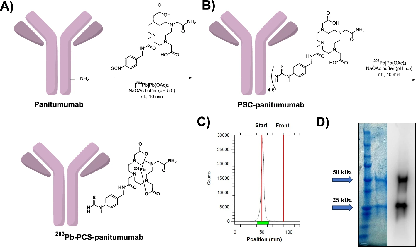
Fig.1. The 203Pb-PSC-panitumumab was produced with 41.5 ± 8% yield, >99% purity, and >95% stability in human serum over 48 hours.
Cellular uptake of 203Pb-PSC-panitumumab in EGFR-expressing FaDu cells increased over time, reaching a plateau at 60 minutes. Specificity was confirmed by blocking studies, showing up to 95% inhibition with increasing panitumumab concentrations, while the immunoreactive fraction was approximately 30%. (Fig.2)
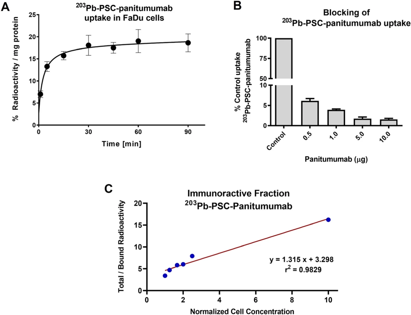
Fig. 2. Cellular uptake and competitive binding studies confirmed specific EGFR targeting of 203Pb-PSC-panitumumab in FaDu cells, supported by Lindmo assay results.
SPECT/CT imaging was performed using the Mediso nanoScan® SPECT/CT system to visualize the biodistribution of 203Pb-PSC-panitumumab in mice bearing EGFR-positive HNSCC tumors, with clear tumor localization observed at both 48 and 120 hours post-injection. Under EGFR blocking conditions, SPECT/CT images demonstrated a significant reduction in tumor uptake accompanied by increased radioactivity in the heart and blood pool, indicating effective receptor blockade. The remaining tumor radioactivity detected despite blocking is attributed to the enhanced permeability and retention (EPR) effect, a phenomenon commonly observed in immunoPET and immunoSPECT imaging of solid tumors. These results highlight the selective and sustained tumor targeting capability of 203Pb-PSC-panitumumab and underscore the utility of this radioligand for specific imaging of EGFR-expressing tumors using the Mediso nanoScan® platform. (Fig.3)
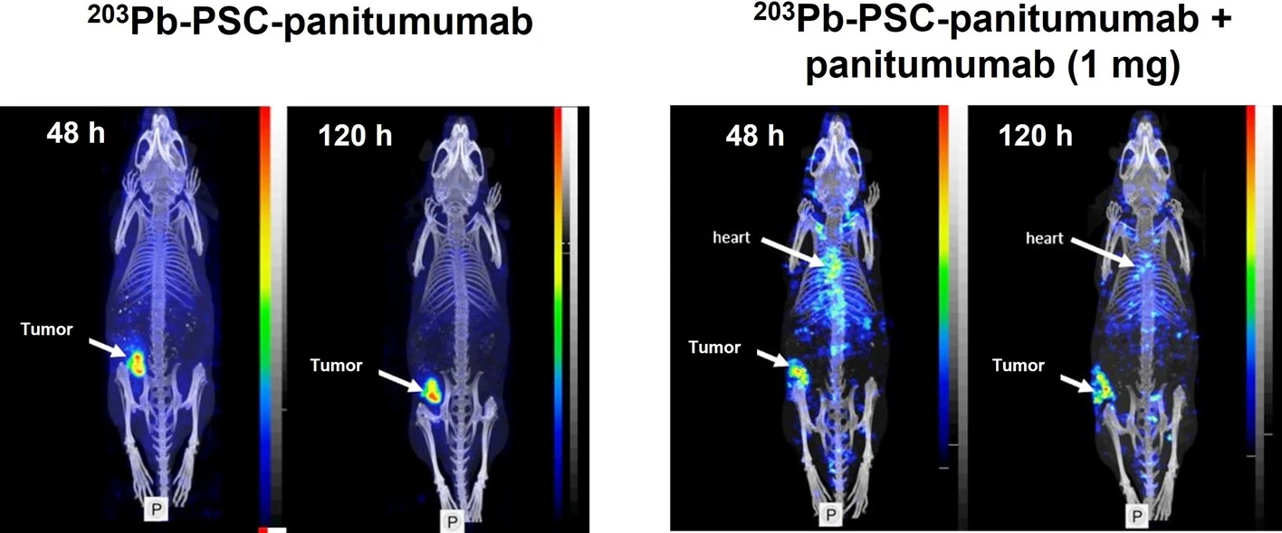
Fig. 3. SPECT/CT images showing NRG mice with EGFR-positive HNSCC PDX tumors at 48 and 120 hours post-injection, comparing control and blocking groups; tumor locations are marked with arrows.
Conclusion
The use of the nanoScan SPECT/CT system enables high-resolution, non-invasive imaging of 203Pb-PSC-panitumumab biodistribution and tumor uptake in vivo, providing precise anatomical localization alongside functional data. Its sensitivity and quantitative capabilities allow for detailed monitoring of EGFR-positive tumors over time, making it an excellent platform for evaluating novel immuno-SPECT probes and supporting the development of personalized radiotheranostic strategies.
Full article on ejnmmipharmchem.springeropen.com


How can we help you?
Don't hesitate to contact us for technical information or to find out more about our products and services.
Get in touch