Augmentation of bone formation by sympathectomy in rats as evaluated by [99mTc]Tc-MDP
2025.07.02.
Zili Cai et al., Frontiers in Endocrinology, 2025
Summary
The role of the sympathetic nervous system in bone metabolism remains unclear. Given that 99mTc-methylene diphosphonate ([99mTc]Tc-MDP) uptake reflects active bone formation and mineralization, this study aims to investigate the effects of sympathetic denervation on bone formation in rats using [99mTc]Tc-MDP SPECT/CT imaging.
Numerous studies have demonstrated that the sympathetic nervous system (SNS) exerts significant regulatory effects on bone formation. However, the role of SNS in bone remodelling remains controversial. While some studies suggest that SNS activation inhibits bone formation via β2-adrenergic receptors, others propose a pro-osteogenic effect under specific physiological conditions. Most existing research has focused on indirect hypothalamic-mediated central nervous system (CNS) regulation of bone metabolism, whereas direct sympathetic regulation of osteogenesis remains poorly characterized. Bone formation is a critical phase of fracture healing, a complex process involving sequential bone resorption and subsequent deposition to replace damaged tissue. Despite its clinical relevance, the interplay between SNS and fracture healing—particularly the balance between osteoblastic bone formation and osteoclastic resorption—has not been thoroughly investigated.
99mTc-methylene diphosphonate ([99mTc]Tc-MDP), a radiopharmaceutical agent widely used in skeletal scintigraphy, provides a functional assessment of bone metabolism by targeting sites of active osteoblastic differentiation and mineralization. Unlike conventional X-ray imaging, which primarily visualizes structural callus formation, [99mTc]Tc-MDP scintigraphy directly correlates with dynamic metabolic activity, offering superior sensitivity for detecting subtle changes in bone turnover. Furthermore, the high sensitivity and specificity of [99mTc]Tc-MDP make it powerful for detecting subtle changes in bone metabolism, which can not be analyzed by Micro-CT. These attributes make [99mTc]Tc-MDP bone scan the only tool available for evaluating the activity of bone formation in vivo following sympathetic denervation.
This study aims to investigate the direct effects of SNS ablation on bone metabolism.
Results from nanoScan® SPECT/CT
A superior cervical ganglionectomy (SCGx) was first performed to achieve localized sympathetic denervation in the cranium, following by establishment of a circular cranial fracture in the form of harvesting a circular bone flap and placing it back in situ.
After all required the procedures, all rats were subjected to Micro SPECT/CT imaging. All rats were injected with [99mTc]Tc-MDP (111 MBq, 500 µL) via the tail vein and imaged using a micro SPECT/CT system (Mediso Medical Imaging Systems, Hungary). CT images were acquired with parameters of 50 kV source voltage, 0.61 mA current, and 300 ms exposure time. Pinhole SPECT images were obtained with the energy peak 140 keV, window width 20%, and a frame rate of 18 frames per second.
Three-dimensional SPECT data were acquired and reconstructed using CT-based attenuation correction and an iterative reconstruction algorithm. The CT component was reconstructed using filtered back-projection. Tissue-associated radioactivity was expressed as SUV. SPECT scans were conducted 60 minutes post-injection with CT images acquired simultaneously. SPECT/CT fusion images were analyzed by nanoScan software (InterView FUSION, Mediso Medical Imaging Systems, Hungary). For the SCGx group and the control group, micro SPECT/CT imaging were performed at 3, 6, 9 weeks, postoperatively. The region of interest (ROI) over the cranium was selected on the transverse slices of the micro SPECT/CT fusion images (Figure 2). The reconstructed images were post-processed with a 3D Gaussian filter and displayed with a slice thickness consistent with SPECT scanning. Bone mineral density (BMD), bone volume (BV), and the ratio of bone volume to tissue volume (BV/TV) were quantified from a defined cylindrical volume of interest (VOI) using CTAn software. A VOI (diameter: 5.4 mm, thickness: 1.2 mm) was selected for analysis. BMD was calculated by converting X-ray attenuation coefficients within the VOI into density values using calibration with hydroxyapatite phantoms. BV and BV/TV were obtained by applying appropriate thresholding to binarize the images, allowing for segmentation of bone tissue, and quantifying the corresponding volume.
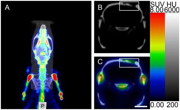
FIGURE 2: Micro SPECT/CT imaging analysis of ROI. (A) Micro SPECT/CT fusion image. (B, C) Micro-CT transverse slice and Micro SPECT/CT fusion transverse slice showing [99mTc]Tc-MDP in the area surrounding the cranial fracture (the rectangular region represents the cranial fracture and the area of [99mTc]Tc-MDP uptake). Scale bar: 5 mm.
Three weeks after SCGx, micro SPECT/CT fusion images showed markedly higher tissue radioactivity in the areas surrounding the cranial fracture of the SCGx group compared to the control group. Nine weeks after SCGx, the control group exhibited consistently low tissue radioactivity in the areas surrounding the fracture. Quantitative analysis revealed [99mTc]Tc-MDP was significantly higher within the area surrounding the fracture of the SCGx group compared to the control group at 3 weeks, 6 weeks, and 9 weeks after SCGx, respectively (Figure 5A). Detailed data were listed in Table 1.

TABLE 1: The uptake of [99mTc]Tc-MDP after SCGx.


FIGURE 5: (A) Representative micro SPECT/CT fusion images. The colour scale represents the level of tissue-associated radioactivity, with red representing the highest uptake, scale bar: 5 mm. (B) The uptake of [99mTc]Tc-MDP after SCGx. (C) Representative images of whole-body SPECT scans. (D) [99mTc] Tc-MDP biodistribution in different skeletal sites. (E) Representative micro-CT and SPECT images. The colour scale represents the level of tissue associated radioactivity, with red representing the highest uptake, scale bar: 5 mm. Red arrows indicate the sagittal suture. (F–H) Summarized data showing the micro-architectural parameters of the newly formed bone tissue by analyzing micro-CT images using image analysis software. BMD (mg/cm³), BV/TV (%) and BV (mm³).
Three weeks after SCGx, micro-CT transverse slices showed bone resorption and blurred shadows surrounding the fracture site in the SCGx group, whereas no significant changes were observed in the control group. Six weeks after SCGx, bone fusion was observed on the side of the fracture line distant from the sagittal suture in the SCGx group, while the fracture line near the sagittal suture appeared blunt. Nine weeks after SCGx, bone fusion was observed near the sagittal suture in the SCGx group, while the control group showed no significant changes in bone formation or resorption. Quantitative micro-CT analysis revealed that the bone histomorphometric parameters (BMD, BV/TV, and BV) were significantly higher in the SCGx group than in the control group at 3, 6, and 9 weeks after SCGx (Figures 5E–H). Detailed data were listed in Table 2.
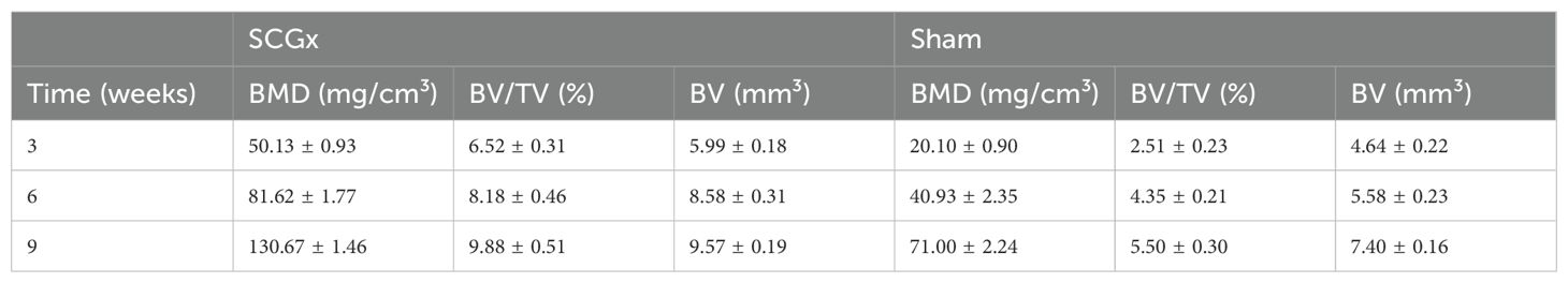
TABLE 2: The bone histomorphometric parameters after SCGx.
Nine weeks after surgery, the lumbar sympathectomy group exhibited significantly higher radioactivity levels compared to the control group in the left femur, right femur, left tibia, right tibia, lumbar vertebrae, and sacrum. However, no significant differences were observed in the cervical vertebrae and thoracic vertebrae between the two groups (Figure 5C). Detailed data were listed in Table 3.
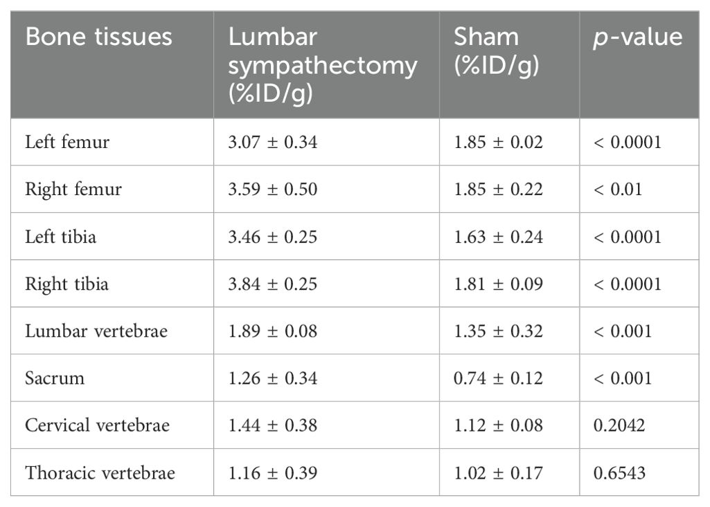
TABLE 3 [99mTc]Tc-MDP biodistribution in different skeletal sites.
Both sympathectomy models revealed significantly enhanced the uptake of [99mTc]Tc-MDP compared to controls. These findings provide novel insights into the regulatory role of SNS in osteogenesis and validate the utility of [99mTc]Tc-MDP as a functional biomarker for bone metabolism assessment.
Conclusion
- Both sympathectomy models revealed significantly enhanced the uptake of [99mTc]Tc-MDP compared to controls.
- SCGx rats exhibited significantly higher [99mTc]Tc-MDP uptake and increased BMD, BV/ TV, and BV in peri-fracture regions at all time points.
- Sympathectomy can enhance osteoblast prolifeation and augment bone formation, which can be effectively assessed and quantified using [99mTc] Tc-MDP SPECT/CT imaging.
- These findings provide novel insights into the regulatory role of SNS in osteogenesis and validate the utility of [99mTc]Tc-MDP as a functional biomarker for bone metabolism assessment.
- This study is the first to utilize [99mTc]Tc-MDP SPECT/CT imaging as a measure to evaluate neurogenic regulation of bone metabolism.
Full article on frontiersin.org


How can we help you?
Don't hesitate to contact us for technical information or to find out more about our products and services.
Get in touch