Implication of 99mTc-sum IL-2 SPECT/CT in immunotherapy by imaging of tumor-infiltrating T cells
2023.02.12.
Yu Gao et al, Journal for ImmunoTherapy of Cancer, 2023
Summary
Tumor-infiltrating T cells, especially activated CD8+ T cells, are vital to immunotherapies, and positively corelate with the prognosis, thus a reliable method for in vivo monitoring of tumor-infiltrating T cells has important clinical values. IL-2-based tracers, such as [18F]FB-IL-2 and 99mTc-IL-2, have been in clinical trials, but the binding affinity of IL-2 to immunosuppressive Treg cells is much stronger than to CD8+ T cells, making them less reliable in assessing immunotherapy responses. In this study, a novel tracer 99mTc-sum IL-2 was developed, which preferentially bound to activated CD8+ T cells rather than Treg cells, therefore, 99mTc-sum IL-2 SPECT/CT showed great potential for predicting immunotherapy response and assessing immunotherapy efficacy in clinical practice. Since non-invasive nuclear imaging can provide real time, in vivo, dynamic, comprehensive, and quantitative information, monitoring the dynamic distribution of T cells in vivo before and during treatment by 99mTc-sum IL-2 SPECT/CT may help to guide the immunotherapy and improve the therapeutic effect for immune checkpoint blockade and adoptive T cell transfer therapy.
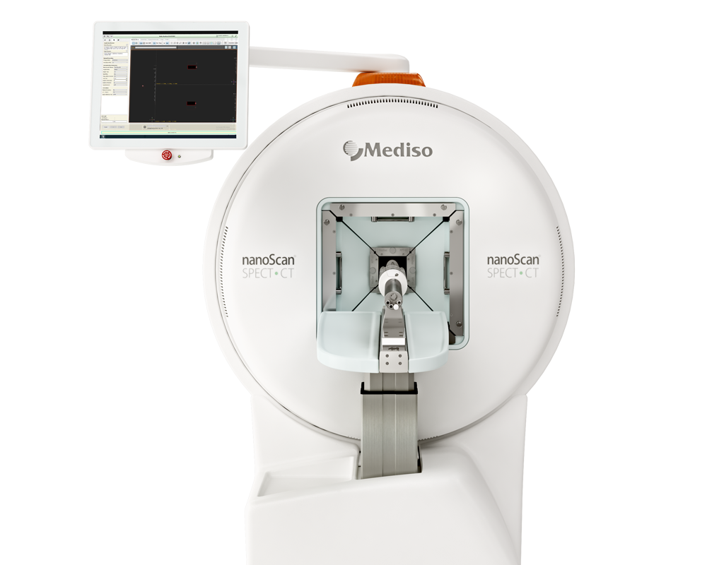
Results from nanoScan® SPECT/CT
For small-animal SPECT/CT imaging, each MC38 tumor bearing mouse was injected via the tail vein with 18.5 MBq of 99mTc-sum IL-2 or 99mTc-IL-2 (n=4). At 0.5hour after injection, mice were imaged using a nanoScan SPECT/CT (Mediso, Hungary) following a standard protocol. The pinhole SPECT images (peak, 140 keV; 20% width; frame time, 25s) were acquired for 13.5min, and CT images were subsequently acquired (50 kVp; 0.67 mA; rotation, 210°; exposure time, 300 ms). All SPECT images were reconstructed and further analyzed with Fusion (Mediso) by drawing volumes of interest on the tumor and major organs.
For the biodistribution study, each MC38 tumor bearing mouse was injected with 0.37 MBq of radiotracer via the tail vein and sacrificed at 0.5hour after injection. Blood, tumors, and organs of interest were harvested, weighed, and counted. The radioactivity in the tissues was measured using a γ-counter. The results are presented as the percentage of injected dose per gram of tissue (%ID/g).
From 1-day post-SPECT/CT scanning, tumor-bearing mice received 12.5mg/kg αPD-L1 antibody treatment, while tumor-bearing mice without treatment were used as controls. One day after the second αPD-L1 treatment, mice were injected with 99mTc-sum IL-2 again for SPECT/CT imaging and biodistribution study, and tumor infiltration of T cells was analyzed by flow cytometry.
Results show:
- SPECT/CT imaging showed strong radioactivity accumulation of 99mTc-sum IL-2 in the MC38 tumors with a clear background at 0.5 h p.i.
- In the blocking imaging, intratumoral injection of excessive sum IL-2 significantly reduced the signal of 99mTc-sum IL-2 in the tumor

Figure 3. 99mTc-sum IL-2 specifically detects T cells in vivo. (A) Representative small animal SPECT/CT images obtained after injection of 99mTc-sum IL-2 in MC38 subcutaneous tumor-bearing mice without or with blocking doses of sum IL-2 protein. (White dashed circle indicates the tumor) (B) Biodistribution of 99mTc-sum IL-2 in MC38 tumor-bearing mice, as well as a blocking study performed by co-injecting 99mTc-sum IL-2 and cold sum IL-2 protein
- Biodistribution study (Figure 3B) confirmed the decrease of tumor uptake at 0.5 hour p.i. suggesting the uptake of 99mTc-sum IL-2 in tumors was specifically mediated. Notably, 99mTc-sum IL-2 was also aggregated in T-cell enriched spleen and tumor-draining lymph node
- As shown in Figure 4 A,B, tumor uptake of 99mTc-sum IL-2 at 0.5-hour p.i. was significantly higher than that of 99mTc-IL-2, indicating that 99mTc-sum IL-2 was more specific than 99mTc-IL-2 to detect tumor-infiltrating T cells in the immune-responsive MC38 tumor model.

Figure 4. (A) Representative small animal SPECT/CT images obtained at 0.5hour after injection of 99mTc-sum IL-2 or 99mTc-IL-2 in MC38 subcutaneous tumor-bearing mice (white dashed circles indicate tumors). (B) Biodistribution of 99mTc-sum IL-2 or 99mTc-IL-2 in MC38 tumor-bearing mice at 0.5h p.i.
- As shown in figure 5B, αPD-L1 treatment markedly increased uptake of 99mTc-sum IL-2 in tumors. Biodistribution study confirmed the significant increase of 99mTc-sum IL-2 uptake in tumors of αPD-L1-treated mice compared with untreated mice, indicating an effective system immunity response promoted by αPD-L1 treatment
- Besides the tumors, αPD-L1 treatment also increased uptake of 99mTc-sum IL-2 in T-cell enriched spleen and TdLN

Figure 5. (B) SPECT/CT imaging of MC38 tumor-bearing mice before and after αPD-L1 treatment. (C) Biodistribution of 99mTc-sum IL-2 in αPD-L1-treated or untreated mice at 0.5h p.i.
Full article on jitc.bmj.com
W czym możemy pomóc?
Skontaktuj się z nami aby uzyskać informacje techniczne i / lub wsparcie dotyczące naszych produktów i usług.
Napisz do nas
