Imaging xCT with Immuno-PET in Therapy-Resistant Non–Small Cell Lung Cancer
2026.06.18.
Abigail R. Barber et al., Journal of Nuclear Medicine, 2026
Summary
Therapy resistance presents a major clinical challenge in non–small cell lung cancer (NSCLC), contributing to its high mortality rate. Overexpression of the amino acid transporter xCT frequently mediates treatment resistance in NSCLC and can be therapeutically targeted with antibody–drug conjugates. The authors developed a corresponding immuno-PET radiotracer to noninvasively evaluate xCT expression in NSCLC and visualize the pharmacokinetics of xCT-targeting antibodies.
Results from nanoScan® PET/CT
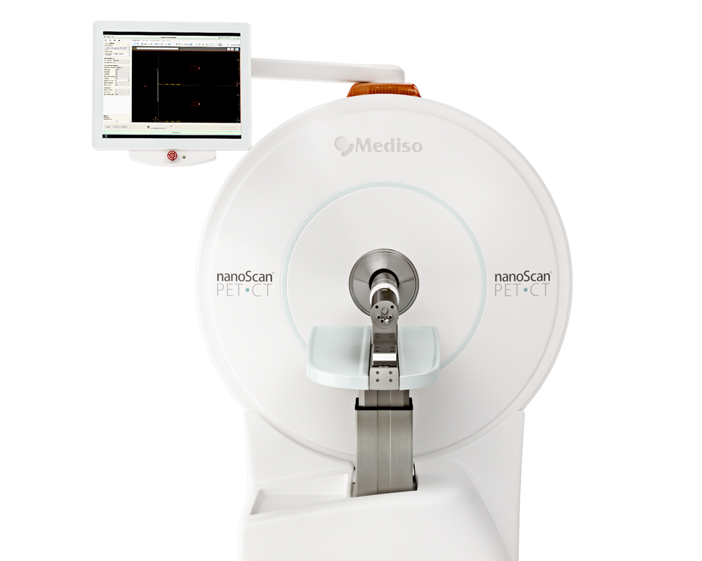
Static PET images were acquired at 4, 24, 48, 72, 96, and 144 h post injection (p. i.) of ~2 MBq [89Zr]Zr-DFO-Abs on a Mediso nanoScan® PET/CT system (1–5 coincidence mode, 3-dimensional reconstruction with CT-based attenuation and scatter corrections) using a 4-bed mouse hotel. Scan time was increased at each time point to account for the half-life of 89Zr (3.3 d), ranging from 30 to 90 min. CT images were acquired for anatomic visualization and attenuation correction (720 projections, helical acquisition, 50 kV, 300 ms exposure). Static PET image reconstruction was performed using the 3-dimensional Tera-Tomo algorithm (4 iterations, 6 subsets, 0.4 mm isotropic voxel size, 400- to 600-keV energy window). InterViewTM FUSION software version 3.11.011 (Mediso) was used for automatic 4-bed hotel separation.
By applying the PET/CT device the two 89Zr-labeled anti-xCT antibodies ([89Zr]Zr-DFO-HM30 and [89Zr]Zr-DFO-HM34) could be evaluated, clearly visualizing xCT-positive H460 NSCLC tumors in vivo. At the first imaging stage both tracers remained predominantly in the blood circulation, tumor uptake became clearly visible from 24 hours onward. Quantification of PET images demonstrated that both antibodies exhibit very similar in vivo pharmacokinetics; no significant difference in tumor uptake could be seen between HM30 and HM34 throughout the imaging period.
 Figure 1. Maximum-intensity-projection (MIP) of [89Zr]Zr-DFO-HM30 and [89Zr]Zr-DFO-HM34 in H460 tumor–bearing mice at 4, 24, 48, 72, and 96 h post injection.
To confirm that tumor accumulation was xCT-specific imaging was performed with [89Zr]Zr-DFO-HM30 in female mice bearing either H1299 (xCT-low) or H460 (xCT-high) subcutaneous tumors. Figure 2 establishes that PET signal depends on target expression rather than passive antibody accumulation by highlighting that H460 (xCT high) tumors are showing strong signal, while H1299 (xCT low) tumors are barely distinguishable from surrounding background.

Figure 2. Representative maximum-intensity-projection (MIP) and axial slice (bottom) PET/CT images of mice bearing subcutaneous H1299 or H460 tumors at 144 h post injection.
Figure 3 evaluates whether the tracer can detect tumors growing inside the lung, a model that better resembles clinical non–small cell lung cancer than subcutaneous xenografts. PET/CT images performed 6 days after tracer injection provides clear visualization of orthotopic H460 lung tumors showing higher tracer uptake in tumor-bearing lungs than in healthy lungs.
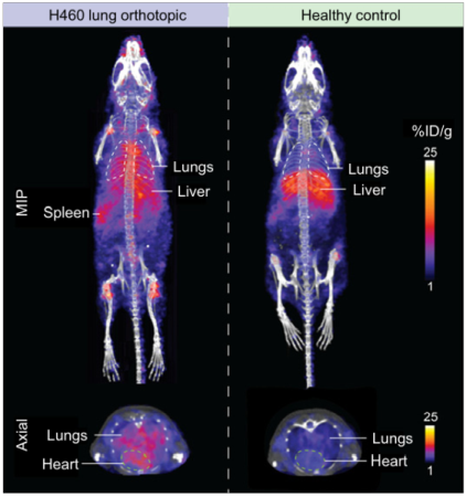
Figure 3. Representative maximum-intensity-projection (MIP, top) and axial slice (bottom) PET/CT images of mice bearing lung orthotopic H460 tumors or healthy control animals at 144 h post injection.
PET imaging also revealed variable uptake among different lung lesions and differences both between separate tumors in the same mouse and between different animals. This indicates that immuno-PET can detect heterogeneity in xCT expression, which is clinically relevant because resistance-associated biomarkers often vary across lesions. Together, these findings position xCT immuno-PET as both an effective imaging tool for drug development and a gateway to theranostic applications in redox-rewired cancers.
Full articel on jnm.snmjournals.org
W czym możemy pomóc?
Skontaktuj się z nami aby uzyskać informacje techniczne i / lub wsparcie dotyczące naszych produktów i usług.
Napisz do nas