Host-directed nanotherapy for the treatment and imaging of tuberculous meningitis
2026.06.10.
Elizabeth W Tucker et al. , Theranostics , 2026
Summary
This study investigated a dendrimer-based nanotherapy (D-NAC) as a new host-directed treatment for tuberculous meningitis (TB meningitis), a severe brain infection in which excessive inflammation causes significant neurological damage.Standard adjunctive corticosteroids often fail to prevent neurological sequelae or improve survival in many populations. Host-directed therapies that can cross the blood-brain barrier (BBB) and reduce neuroinflammation are urgently needed.
The study demonstrates that dendrimer-NAC (D-NAC) can serve as both a diagnostic tool to identify neuroinflammation and a targeted treatment to reduce inflammation and brain injury.
Results from nanoScan® PET/CT
PET imaging showed that D-NAC reached inflammatory brain lesions, reduced neuroinflammation, and improved BBB integrity in rabbits with TB meningitis
- ¹²⁴I-dendrimer PET demonstrated selective accumulation in TB meningitis brain lesions, primarily localizing to activated microglia, the main inflammatory cells involved in TB meningitis
- ¹²⁴I-DPA-713 PET showed that D-NAC treatment reduced microglial/macrophage activation
- ¹⁸F-py-albumin was used to assess blood-brain barrier (BBB) permeability. The results showd improved blood-brain barrier (BBB) integrity following D-NAC treatment.
- ¹⁸F-FDG PET was used to monitor cerebral glucose metabolism longitudinally as part of the treatment assessment.
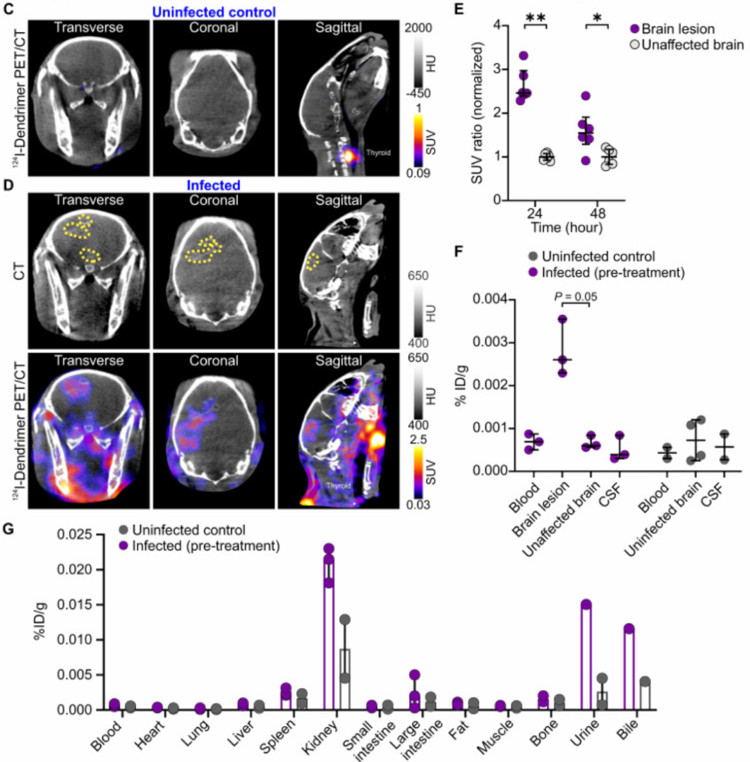
Figure 2. (C and D) Representative transverse, coronal and sagittal 124I-dendrimer PET/CT imaged 24 hours (h) post-tracer injection and three weeks after injection with (C) PBS (uninfected control) or (D) M. tuberculosis (infected). The brain lesion hyperdensity (yellow dotted outline) was seen on CT (D, upper panel) and co-localized with 124I-dendrimer PET signal (D, lower panel) in the infected rabbit. (E) Serial 124I-dendrimer PET imaging presented standard uptake value (SUV) ratio (normalized to the mean of unaffected brain SUV) at 24 and 48 h post-tracer injection in infected rabbits. Each dot represents a volume of interest (VOI). (F and G) Post-mortem ex vivo biodistribution of 124I-dendrimer 48 h post-tracer injection using gamma counting (% injected dose [ID]/g) in the (F) CNS and (G) other organs/bodily fluids.
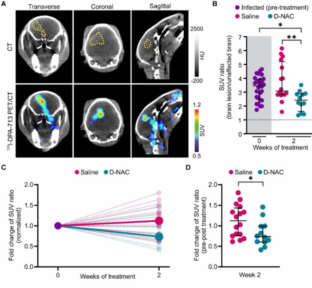
Figure 4. D-NAC decreases microglial activation and density. (A) Representative transverse, coronal, and sagittal 124I-DPA-713 PET/CT images of an infected rabbit imaged three weeks post-infection before treatment. The brain lesion (yellow dotted outline) is visible on CT (upper panel) and co-localizes with 124I-DPA-713 signal (lower panel). (B) PET-derived SUV ratios (brain lesion/unaffected brain) over treatment duration. Each dot represents an individual VOI. (C) PET-derived SUV ratio fold change normalized to week 0, shown for individual VOIs (small, light circles) and group medians (large, dark circles). (D) Comparison of SUV ratio fold change to pre-treatment levels for individual VOIs after two weeks of treatment.
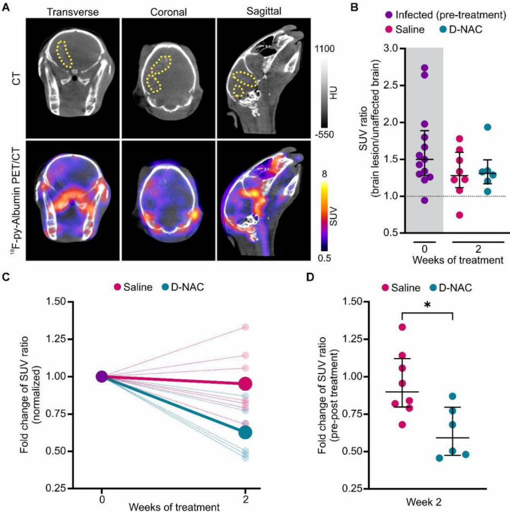
Figure 6. 18-F-Py-Albumin demonstrates healing BBB with D-NAC. (A) Representative transverse, coronal, and sagittal 18F-py-albumin PET/CT images of an infected rabbit imaged three weeks post-infection before treatment. The brain lesion (yellow dotted outline) is seen on CT (upper panel) and co-localizes with 18F-py-albumin signal (lower panel). (B) PET-derived SUV ratios (brain lesion/unaffected brain) over treatment duration. (C) PET-derived SUV ratio fold change normalized to week 0, shown for individual VOIs (small, light circles) and group medians (large, dark circles). (D) Comparison of SUV ratio fold change to pre-treatment levels for individual VOIs after two weeks of treatment.

W czym możemy pomóc?
Skontaktuj się z nami aby uzyskać informacje techniczne i / lub wsparcie dotyczące naszych produktów i usług.
Napisz do nas