Imaging and targeting S1PR1 in HER2+ tumors
2025.10.15.
Shayla Shmuel et al., Nuclear Medicine and Biology, 2025
Summary
Human Epidermal Growth Factor Receptor 2 (HER2) is a membrane receptor tyrosine kinase overexpressed in a subset of gastric cancers and is the target of multiple clinically approved therapies, including the antibody-drug conjugate trastuzumab deruxtecan (T-DXd). However, resistance to HER2-directed therapies remains a major challenge in gastric cancer. Sphingosine-1-phosphate receptor 1 (S1PR1), a G-protein-coupled receptor involved in oncogenic signaling, has been associated with poor prognosis and therapy resistance. This study investigated the role of S1PR1 in modulating response to HER2-targeted therapy and explored therapeutic dual targeting of HER2 and S1PR1. We used immunohistochemistry, Western blot analyses, and S1PR1-targeted radiotracer to assess S1PR1 expression in patient-derived xenograft samples and preclinical tumor models. We observed decreased S1PR1 in tumors that responded to T-DXd and trastuzumab therapy. In contrast, tumors with persistent S1PR1 expression exhibited resistance to HER2-targeted therapy. The S1PR1 inhibitor fingolimod, when combined with T-DXd, significantly enhanced therapeutic efficacy in HER2+/S1PR1+ tumors, resulting in reduced HER2 and S1PR1 protein levels and decreased tumor volume. This work demonstrates that S1PR1 expression is associated with resistance to HER2-targeted therapy, and S1PR1 PET has potential as a biomarker for selecting patients for HER2-targeted therapy, and S1PR1 has the potential to monitor T-DXd therapeutic response.
Results from nanoScan® PET/CT
- The authors used a S1PR1-targeted F18-FS1P1 and C-11-CS1P1 radiotracer to image S1PR1 expression in preclinical tumor models.
- PET imaging was able to quantitatively measure changes in S1PR1 expression in tumors under different treatment conditions
- Tumors that responded to HER2-targeted therapies showed decreased S1PR1 signal / tracer uptake, whereas tumors with persistent S1PR1 expression showed resistance to those therapies.
- The data support the use of S1PR1 PET as a biomarker for selecting tumors likely to respond to HER2 therapy, and for monitoring therapeutic response.
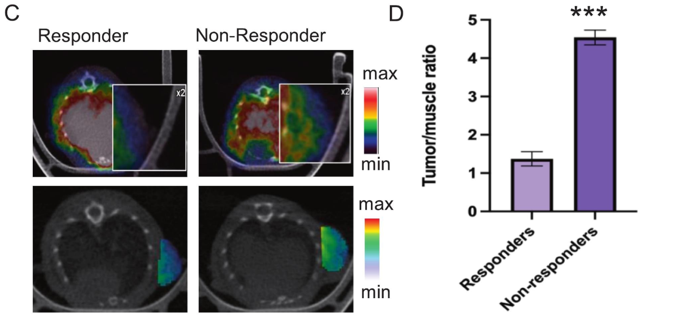
Figure 3 C) [18F]FS1P1 PET/CT images of T-DXd responders and non-responders. PET/CT images at the bottom show the tumor signal only. The tumor was extracted by using the shape from the CT to outline the tumor, and then, once the CT was extracted, the CT image was overlaid on the PET signal in the tumor. D) Tumor-to-muscle ratio of [18F]FS1P1 in T-DXd responders and non-responders.

Hogyan segíthetünk Önnek?
További termékinformációkért, vagy támogatásért keresse szakértőinket!
Vegye fel a kapcsolatot