Targeted PET Imaging of Pseudomonas aeruginosa Using a [68Ga]Ga-Pyoverdines Cocktail
2026.08.13.
Kristyna Krasulova et al., Journal of Nuclear Medicine, 2026
Summary
Pseudomonas aeruginosa is a major cause of nosocomial infections. Current diagnostic methods often lack sensitivity and specificity, resulting in a delayed or inaccurate diagnosis. To address these limitations, new diagnostic strategies are being developed. Molecular imaging techniques, such as PET, allow localization and monitoring of infections in situ; however, specific radiotracers for P. aeruginosa imaging are lacking. Radiolabeled siderophores have shown promise as diagnostic tools for PET imaging. Pyoverdines, specific siderophores produced by P. aeruginosa, are classified into 3 types: pyoverdine type I (PVDI), pyoverdine type II (PVDII), and pyoverdine type III (PVDIII). In this study, the authors evaluated a radiotracer specifically targeting P. aeruginosa infections using radiolabeled pyoverdines.
Results with nanoScan PET/MRI® 3T and PET/CT®
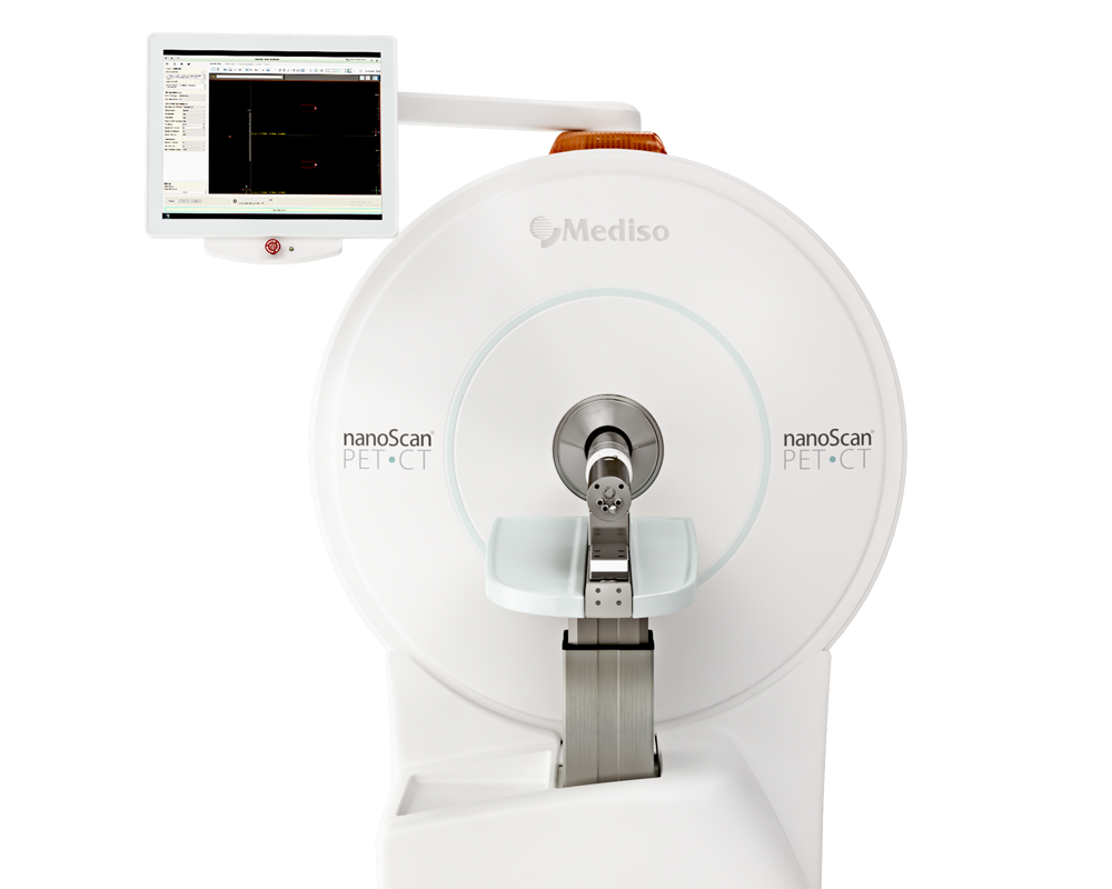
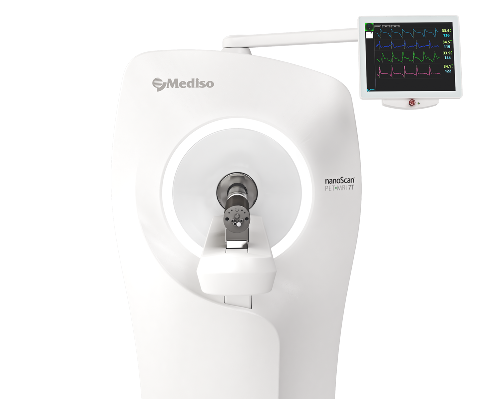
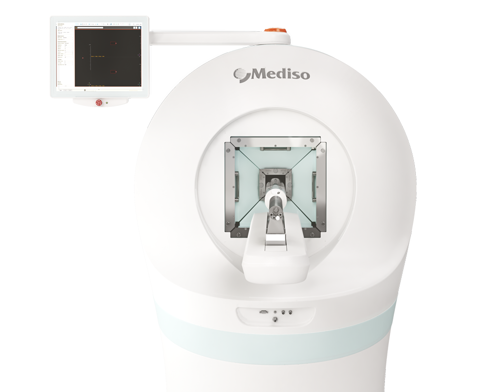
Female BALB/c mice and Lewis rats age 8–10 wk (Envigo) were used in the experiments. Animals were injected with 5-10 MBq of [68Ga]Ga-PVDI, [68Ga]Ga-PVDII, [68Ga]Ga-PVDIII or [68Ga]Ga-pyoverdines cocktail (via the tail vein) and placed in the prone position in a nanoScan PET/CT or nanoScan PET/MRI 3T system (Mediso Medical Imaging Systems, Hungary). After the administration of [68Ga]Ga-labeled tracers, static PET imaging was initiated at 45 min post injection, followed by whole-body helical CT or MRI scans. The final images were normalized to the injected activity and animal weight, with the results presented on an SUV scale.
-
The fused PET/MR images clearly localized tracer accumulation in the infected left hind leg, while the saline-injected contralateral leg showed no relevant uptake as it can be seen on Figure 1. Importantly, each pyoverdine preferentially accumulated in infections caused by the corresponding aeruginosa siderotype: [68Ga]Ga-PVDI for strain ATCC15692, [68Ga]Ga-PVDII for ATCC9027, and [68Ga]Ga-PVDIII for ATCC15392. The PET/MRI images therefore provide both anatomical localization and functional information, demonstrating the ability of targeted PET to distinguish different P. aeruginosa strains in vivo. Quantitative analysis of the PET signal confirmed significantly higher tracer uptake in infected compared with control sites.
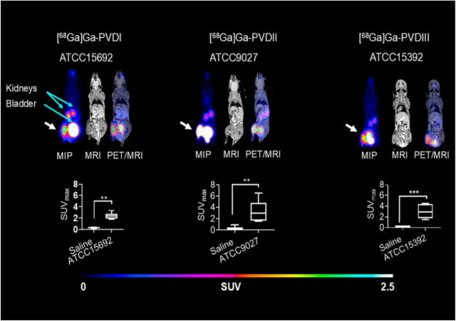
Fig. 1. Maximum-intensity-projection (MIP), MR, and fused PET/MR sagittal slice images of mice with P. aeruginosa infection (strains ATCC15692, ATCC9027, ATCC15392—each representing 1 of 3 groups) in left hind leg (white arrows) and saline injection in right hind leg.
-
The fused PET/MR images on Figure 2. show pronounced tracer accumulation at the P. aeruginosa-infected sites, whereas no relevant radioactive signal was detected in the sites containing S. aureus or sterile inflammation. This result highlights the advantage of combining molecular PET imaging with high-resolution MR anatomy: the infection sites can be accurately localized while the PET component provides pathogen-specific functional information. The findings confirm that [68Ga]Ga-PVDI, [68Ga]Ga-PVDII, and [68Ga]Ga-PVDIII selectively target the corresponding P. aeruginosa infections rather than nonspecific inflammatory processes.
Fig. 2. Maximum-intensity-projection (MIP) and fused PET/MR sagittal slice images with P. aeruginosa ATCC15692 (A) ATCC9027 (B) and ATCC15392 (C) in left hind leg (white arrows) and S. aureus (orange arrows) in right hind leg (mouse 1) and sterile inflammation in right hind leg (yellow arrows) (mouse 2).

-
The applicability of the [68Ga]Ga-pyoverdines for imaging a clinically relevant pulmonary infection was investigated in an immunosuppressed rat model. The PET/CT maximum-intensity-projection images on Figure 3. demonstrate focal tracer accumulation in the lungs of infected animals. Each individual tracer showed uptake corresponding to the P. aeruginosa strain-specific pyoverdine system, while the cocktail enabled detection across the tested strains. The combination of PET with CT provided functional visualization of the radiotracer uptake together with anatomical localization of the pulmonary infection.
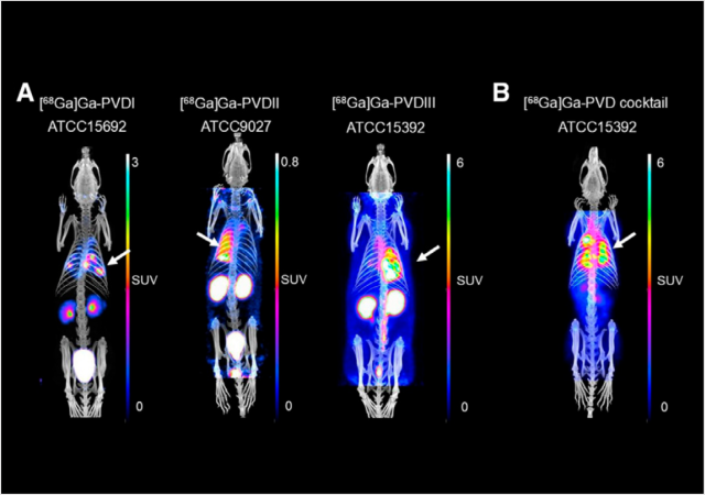
Fig. 3. PET/CT maximum-intensity-projection images of [68Ga]Ga-pyoverdines in rat lung model of P. aeruginosa infection (24 h after infection and 45 min post injection of [68Ga]Ga-PVDI (ATCC15692), [68Ga]Ga-PVDII (ATCC9027), [68Ga]Ga-PVDIII (ATCC15392) (A), and [68Ga]Gapyoverdines cocktail (ATCC15392) (b). Immunosuppressed rats received 100 mL of suspension containing (2–5) 3 108 viable cells/mL. White arrows indicate site of infection.
-
PET/MRI was used to further investigate whether the [68Ga]Ga-pyoverdines cocktail specifically targets viable aeruginosa. Mice were inoculated with viable bacteria in one hind leg and heat-inactivated bacteria in the contralateral leg, followed by a scan 45 min post injection. The MIP images and fused PET/MR slices confirm tracer accumulation at sites containing viable P. aeruginosa, whereas the sites containing heat-inactivated bacteria showed no relevant radioactive signal (see Figure 4.). Quantitative SUVmax analysis also reinforced the statistically significant difference between viable and heat-inactivated bacterial sites. The PET/MRI results therefore highlight that the imaging signal is associated with active bacterial infection rather than merely with the presence of bacterial material.
Fig. 4. Maximum-intensity-projection (MIP) PET image, sagittal slices of MR, and fused PET/MR images of mice infected with P. aeruginosa ATCC15692 (A) ATCC9027 (B), and ATCC15392 (C) in left hind leg (white arrows) and corresponding heat-inactivated P. aeruginosa strains (red arrows) in right hind leg. (D) Comparison of quantification of radioactive signal uptake among left hind legs injected with viable P. aeruginosa strains and right hind legs injected with heat-inactivated P. aeruginosa strains.

-
Figure 5. illustrates the sensitivity of the [68Ga]Ga-pyoverdines cocktail for detecting pulmonary P. aeruginosa infections at substantially lower bacterial loads. Compared with the healthy control, focal tracer accumulation was clearly visualized in the lungs of animals infected with ATCC15692, ATCC9027, and ATCC15392, despite the lower infectious doses used in this experiment. The fused PET/CT images combine the functional information provided by the targeted [68Ga]Ga-pyoverdines with the anatomical information from CT, enabling direct localization of the pulmonary infection sites. The study reports that the cocktail successfully detected all tested P. aeruginosa strains even at infection doses in the range of 10⁵–10⁶ viable bacterial cells, revealing the potential of PET/CT for sensitive and noninvasive detection of low-burden bacterial infections.
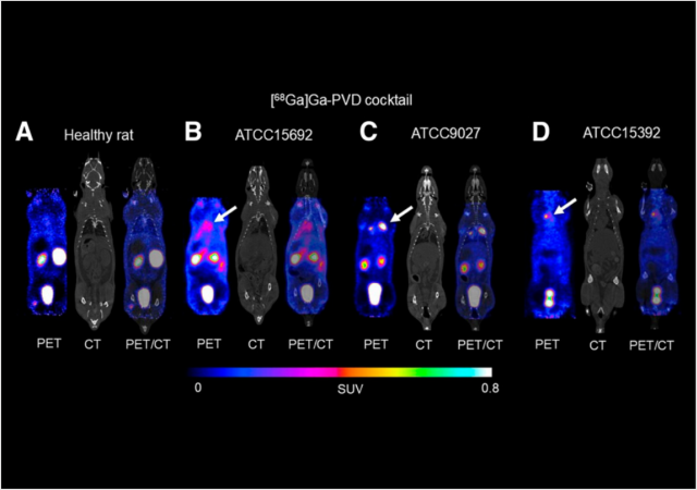
Fig. 5. PET, CT and fused PET/CT coronal slices of images of [68Ga]Ga-pyoverdines cocktail in rat lung infection model. Immunosuppressed rats were infected with significantly lower infectious dose (compared with Fig. 5). (A) and in lungs infected with ATCC15692 (B), ATCC9027 (C), and ATCC15392 (D). White arrows mark infection sites.
Full article on jnm.snmjournals.org
¿como podemos ayudarlo?
Póngase en contacto con nosotros para obtener información técnica, productos y servicios!
Ponerse en contacto