Myoglobin-loaded gadolinium nanotexaphyrins for oxygen synergy and imaging-guided radiosensitization therapy
2023.10.04.
Xiaotu Ma et al, Nat Commun. 2023
Summary
This study explores the enhancement of gadolinium-coordinated texaphyrin (Gd-Tex) radiosensitization through self-assembling nanovesicles, termed Gd-nanotexaphyrins (Gd-NTs). By incorporating myoglobin (Mb), the resulting Mb@Gd-NTs alleviate tumor hypoxia, significantly improving Gd-Tex radiosensitization. The versatile nanoplatform can also chelate 177Lu3+ (Mb@177Lu/Gd-NTs) for dual-modality imaging. This "one-for-all" nanoplatform demonstrates potential for imaging-guided radiosensitization therapy with broad clinical applications.Researchers develop gadolinium nanotexaphyrins (Gd-NTs) that, through self-assembly and myoglobin loading, enhance radiosensitization efficacy of Gd-coordinated Texaphyrin, alleviating tumor hypoxia and suppressing recurrence in radiotherapy.Radiotherapy (RT) is effective but faces limitations, including potential damage to normal tissues and recurrence. Gadolinium-coordinated texaphyrin (Gd-Tex) was a promising radiosensitizer, but its efficacy was limited. Researchers transformed Gd-Tex into Gd-nanotexaphyrins (Gd-NTs), nanovesicles with lipid bilayers, significantly improving tumor accumulation and radiosensitization efficacy, addressing a key challenge in radiotherapy.

Fabrication and characterization of Mb@177Lu/Gd-NTs.
a Diagram of the fabrication of Mb@177Lu/Gd-NTs. b Illustration of imaging-guided radiosensitization therapy. Intravenous injection of Mb@177Lu/Gd-NTs enables SPECT/MRI dual-modality imaging for accurately monitoring drug delivery in real-time. Mb@177Lu/Gd-NTs can significantly increase Gd-Tex accumulation in tumors and obviously relieve tumor hypoxia. O2 synergically enhances the radiosensitization effect of Gd-Tex, leading to the decreased reductants and increased ROS in tumor cells, causing the enhanced cell apoptosis, eventually inducing long-term immunological antitumor memory. c, d TEM images (c) and hydrodynamic diameter distribution (d) of Gd-NTs (I) and Mb@Gd-NTs (II). Scale bar, 100 nm. These experiments (c, d) were repeated three times independently with similar results. e The UV–Vis absorption spectra of Tex-lipid, Gd-Tex-lipid and Gd-NTs. The experiment (e) was repeated three times independently with similar results. f The concentration of dissolved oxygen released from different concentrations of MbO2@Gd-NTs (0-80 μg/mL Mb). g The concentration of dissolved oxygen released from free Hb, HbO2@NTs, free Mb, and MbO2@NTs (40 μg/mL Hb or Mb). MbO2, oxygenated Mb. HbO2, oxygenated Hb. These experiments (f, g) were repeated three times independently with similar results. h The kinetics of ascorbate oxidation catalyzed by NTs, MbO2@NTs, Gd-NTs, MbO2@Gd-NTs under hypoxic conditions. The data (h) are shown as the mean ± SD (n = 3 independent experiments). Source data are provided as a Source Data file.
The radiosensitization efficacy of Gd-Tex is enhanced by loading myoglobin into Gd-NTs (Mb@Gd-NTs), relieving tumor hypoxia and improving Gd-Tex radiosensitization. The nanoplatform, capable of chelating Gd3+ for MRI and 177Lu3+ for SPECT imaging, allows real-time tracking of drug delivery, demonstrating potential for imaging-guided radiosensitization therapy with broad clinical applications.
Materials and methods
Materials were sourced as follows: Gadolinium acetate hydrate and skeletal muscle Mb were from Sigma–Aldrich (USA), triethylamine from Alfa Aesar (USA), and lipid components (DSPC, DSPE-mPEG2000, cholesterol) from Avanti Polar Lipids (USA). Cell culture items were purchased from Corning (USA).Immunological assay kits included the Mouse IFN-γ ELISA Kit (CZM10-96) from Beijing CHENG ZHI KE WEI Biotechnology Co., Ltd. (China) and the Mouse IL-1β ELISA Kit (EMC001bQT) from NeoBioscience Technology Co., Ltd. (China). Ultrapure water was obtained through the Millipore Milli-Q Gradient System (USA).Antibodies for protein detection and flow cytometry were procured from different suppliers: Rabbit anti-γ-H2A.X (phospho S139) antibody and FITC-conjugated goat anti-rabbit IgG H&L antibody from Abcam (USA). Flow cytometry antibodies were purchased from BioLegend (USA).Mice were randomly assigned groups based on pre-administration bioluminescent imaging, ensuring comparable average tumor sizes. In vivo tracking of nanovesicle behavior utilized SPECT, MRI, and fluorescence imaging, with quantification of signal intensity in the region of interest (ROI) through respective software. Each group comprised a minimum of three mice for robust statistical analysis, and investigators conducting the animal experiments were not blinded to group information.
Results from nanoScan SPECT/CT
SPECT/CT imaging and biodistribution studies of Mb@177Lu/Gd-NTs
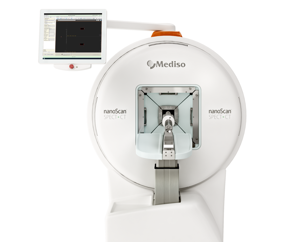
- Small animal SPECT/CT imaging of LLC tumor-bearing mice was performed using a nanoScan® SPECT/CT system (Mediso Ltd, Hungary).
- Mice, each receiving an intravenous injection of 400 μCi Mb@177Lu/Gd-NTs, underwent SPECT/CT imaging at 2, 12, 24, 48, and 72 h post-injection. SPECT images were reconstructed using an OSEM algorithm, and fusion images with cone beam CT were generated. Biodistribution studies on LLC tumor-bearing mice (tumor volume ~100 mm3) involved injecting 50 μCi of Mb@177Lu/Gd-NTs, followed by sacrifice at 6, 24, 48, and 72 h post-injection.
- Tissue samples were collected, weighed, and their radioactivities were measured to calculate the percentage of injected dose per gram of tissue (%ID/g) using an automatic γ-counter.
Single-photon emission computed tomography (SPECT) and magnetic resonance imaging (MRI) bimodal imaging, pharmacokinetics and biodistribution studies
- Investigating Mb@Gd-NTs for MRI contrast, we assessed their in vitro effect at 7 T, revealing a significant enhancement with an r1 value of 0.62 mM−1 S−1.
- Surfactant-dissociated Mb@Gd-NTs exhibited a higher r1 value (0.77 mM−1 S−1), suggesting improved contrast after endocytosis in tumor cells.
- Intravenous administration of Mb@Gd-NTs notably increased the MRI signal in the tumor region, peaking at 24 h post-injection, indicating substantial accumulation within the tumor.

MRI/SPECT/CT imaging, pharmacokinetics and biodistribution of Mb@Gd-NTs.
a In vitro phantom MRI contrast images of different concentrations of Mb@Gd-NTs. b Linear fitting of R1 (1/T1) values as a function of different concentrations of Gd-Tex-lipid and the r1 value of the intact or dissociated Mb@Gd-NTs. c In vivo T1-weighted MRI imaging of LLC tumor-bearing mice before and 4 h, 24 h, and 48 h after the injection of Mb@Gd-NTs. d SPECT/CT images of LLC tumor-bearing mice acquired at 2–72 h after i.v. injection of Mb@177Lu/Gd-NTs. These experiments (a–d) were repeated three times independently with similar results. e Biodistribution of Mb@177Lu/Gd-NTs in tumors and major organs measured at 6, 24, 48, and 72 h post-injection. The data (e) are shown as the mean ± SD (n = 4 mice). f In vivo fluorescence imaging of LLC tumor-bearing mice 0.5–72 h after i.v. injection of free Cy5.5-Mb, Cy5.5-Mb@NTs and Cy5.5-Mb@Gd-NTs (n = 4 mice, cf. Supplementary Fig. 9). g The content of Cy5.5- Mb or Cy5.5-Hb in lungs, muscles, and tumors 2 h after the injection of free Cy5.5-Mb and 24 h after the injection of Cy5.5-Mb@NTs, Cy5.5-Hb@NTs, and Cy5.5-Mb@Gd-NTs. The data (g) are shown as the mean ± SD (n = 4 mice). h Pharmacokinetic studies of Cy5.5-labeled different agents. The data (h) are shown as the mean ± SD (n = 4 mice). i, j Immunofluorescent staining of the hypoxic region (pseudo-red color) of tumor tissue sections and the corresponding semi-quantification results (j). Cell nuclei were stained with DAPI (pseudo-blue color). Tumors were excised from mice 2 h after the injection of free Mb and 24 h after the injection of different liposomes. Scale bar, 60 μm. The data (j) are shown as the mean ± SD (n = 6 mice). Statistical analysis was performed by a two-tailed unpaired t test (g, h, j). *P < 0.05; **P < 0.01; ***P < 0.001. Source data are provided as a Source Data file.
- Radiolabeled Mb@Gd-NTs with 177Lu for SPECT imaging demonstrated clear tumor visualization, reaching peak uptake at 24 h post-injection, consistent with MRI findings.
- Biodistribution revealed significant accumulation in tumors, liver, and spleen, typical of nanoparticle behavior.
- Tumor uptake of Mb@Gd-NTs was highest at 24 h (6.92 %ID/g). Fluorescence imaging with Cy5.5-labeled Mb@Gd-NTs showed superior tumor retention compared to free Cy5.5-Mb.
- Quantification confirmed a 7.88-fold higher tumor uptake with Mb@Gd-NTs, attributed to prolonged blood circulation. Pharmacokinetics studies indicated significantly longer half-lives for Mb@Gd-NTs compared to free Mb.
- Mb@Gd-NTs effectively alleviated tumor hypoxia, outperforming Hb@Gd-NTs. The results suggest the potential of Mb@Gd-NTs for imaging-guided dosimetry in treatment planning.
Incorporating Mb into the nanoplatform extended its half-life and enhanced its effectiveness in alleviating hypoxia. This versatile and biocompatible nanoplatform, featuring radiosensitization and SPECT/MRI imaging capabilities, holds great promise for clinical applications in imaging-guided radiotherapy.
Full article ncbi.nlm.nih.gov
¿como podemos ayudarlo?
Póngase en contacto con nosotros para obtener información técnica, productos y servicios!
Ponerse en contacto