GRPR-Antagonists Carrying DOTAGA-Chelator via Positively Charged Linkers: Perspectives for Prostate Cancer Theranostics
2024.04.08.
Karim Obeid et al, Pharmaceutics, 2024
Summary
Gastrin-releasing peptide receptor (GRPR)-antagonists have been used as motifs in the development of theranostic radioligands for prostate cancer. GRPR is highly expressed in early-stage prostate cancer with advantageously no expression in benign prostatic hyperplasia. On the other hand, GRPR expression patterns become less consistent in metastatic advanced stages of the disease. Numerous theranostic radiopharmaceuticals with promising preclinical results for use in the management of prostate cancer have been developed over the years based on GRPR-antagonists. Antagonists are the preferred option for injection into patients because they do not activate the GRPR upon binding and hence do not elicit acute adverse effects. Furthermore, radiolabeled GRPR-antagonists have shown superior pharmacokinetic profiles compared with agonists in animal models and humans.
There have been developed a number of RM2-related analogs as radiopharmaceutical candidates for prostate cancer theranostics, carrying different types of radiometal chelators via diverse linkers. To implement targeted therapy, the DOTAGA (1,4,7,10- tetrakis(carboxymethyl)-1,4,7,10-tetraazacyclo-dodecane glutaric acid) chelator has been introduced in several analogs, known to form stable complexes with a number of therapeutic radiometals, including beta (Lu-177, Y-90) and alpha emitters (Ac-211, Bi-213).
The prospects of AU-RM26-M2/3/4, seen together as theranostic In-111/Lu-177 pair candidates (for SPECT diagnostic imaging/radionuclide therapy), were first assessed in the In-111 analogs and in comparison with the [111In]In-AU-RM26-M1 reference. In this way, the best performing compound(s) could be selected for future evaluation in combination with the respective [177Lu]Lu-labelled therapeutic counterpart(s). Unsurprisingly, incorporation of In-111 by DOTAGA was successful for all new peptide conjugates, resulting in high-quality and high-purity radioligands. The [111In]In-DOTAGA radiometalchelate was found to be stable both in human serum and during challenge conditions.
These studies have been focused on the development of radiolabelled RM26 (H-DPhe6–Gln7–Trp8–Ala9–Val10–Gly11–His12–Sta13–Leu14–NH2) analogs, such as [111In]In-DOTAGA-PEG2-RM26. The improved metabolic stability (resistance to degradation by neprilysin (NEP)) and enhanced tumor uptake in mice of [111In]In-DOTAGA-PEG2-[Sar11]RM26 ([111In]In-AU-RM26-M1) in a head-to-head comparison study with unmodified [111In]In-DOTAGA-PEG2-RM26 has been recently demonstrated.
Aiming at further improvements of the pharmacokinetic profile of [111In]In-AU-RM26-M1, the efforts were next directed toward prolonging tumour retention and increasing clearance from healthy tissues, especially from excretory organs (like the kidneys), thereby maximizing the therapeutic index. For this purpose, three new [111In]In-AU-RM26-M1 analogs, carrying different types and numbers of basic residues in the linker were introduced.
These three new In-111-labelled AU-RM26-M1 mimetics with base residues in the linker are presented as follows: (i) AU-RM26-M2 (PEG2-Pip), (ii) AURM26-M3 (PEG2-Arg) and (iii) AU-RM26-M4 (Arg-Arg-Pip). The biological profile of the new bioconjugates after labelling with In-111 was evaluated and compared in GRPR-expressing prostate adenocarcinoma PC-3 cells and animal models vs. AU-RM26-M1 (reference) and their potential discussed as radiotherapeutic candidates in prostate cancer after labelling with particle emitting radiometals, like Lu.
Results from nanoScan® SPECT/CT
The BALB/C nu/nu mice were implanted with GRPR-positive prostate cancer xenografts by subcutaneous injection on the right hind leg of PC-3 cells suspended in PBS four weeks before the biodistribution studies.
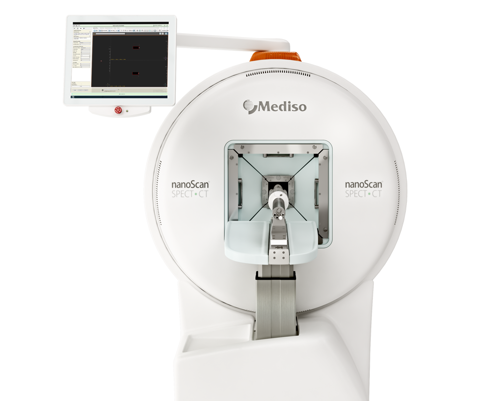
SPECT/CT imaging was performed in two different mice for [111In]In-AU-RM26-M2 and [111In]In-AU-RM26-M4 both at 4 h pi (under anaesthesia) and at 24 h pi (after euthanasia with CO2); a bolus of the radioligand was iv injected in each mouse. Whole body scans were performed using nanoScan SPECT/CT (Mediso Medical Imaging Systems, Budapest, Hungary). The acquisition time was 20 min. SPECT raw data was reconstructed using Tera-TomoTM 3D SPECT reconstruction technology (version 3.00.020.000; Mediso Medical Imaging Systems Ltd., Budapest, Hungary). CT data was reconstructed using Filter Back Projection and fused with SPECT files using Nucline 2.03 Software (Mediso Medical Imaging Systems Ltd., Budapest, Hungary).
 Figure 8. Comparative SPECT/CT images of mice bearing PC-3 xenografts for [111In]In-AU-RM26-M2 at (a) 4 h and (b) 24 h pi and for [111In]In-AU-RM26-M4 at (c) 4 h and (d) 24 h pi; green arrows are directed toward the kidneys and orange arrows at the implanted PC-3 tumours; scans were presented as maximum intensity projections in the red/green/blue colour scale.
Concordant with biodistribution results, the PC-3 tumours and the kidneys were visualized at both time points against a clear background. [111In]In-AU-RM26-M4 displayed higher tumour uptake and lower renal radioactivity compared with [111In]In-AU-RM26-M2 at both time intervals. The type of positively charged residues in the linker of AU-RM26-M1 mimics strongly influenced biological behaviour.
- Three [111In]In-AU-RM26-M1 mimics carrying the [111In]In-DOTAGA radiometal-chelate at the N-terminal DPhe6 of [Sar11]RM26 through positively charged linkers were compared.
- The new analogs showed high affinity and specificity for the GRPR, exhibiting an uptake and distribution pattern in PC-3 cells typical for a radiolabelled GRPR-antagonist.
- Both the type and the number of basic residues in the linker were shown to exert a profound impact on a series of important biological features of the new analogs, such as receptor affinity, uptake and internalization in PC-3 cells, in vivo stability and biodistribution patterns in mice models.
- [111In]In-AU-RM26-M4 with an Arg-Arg-Pip triplet as a linker displayed the highest tumour uptake, while requiring longer times to clear from the background.
- [111In]In-AU-RM26-M2 (PEG2-Pip linker) showed faster background clearance, albeit lower tumour uptake in mice.
- The type of positively charged residues in the linker of AU-RM26-M1 mimics strongly influenced biological behaviour.
- The analogs with Pip next to DPhe6 demonstrated the best overall characteristics and warrant further investigation.
- The validity of these results should be next confirmed for the Lu-177 therapeutic counterparts to establish the applicability prospects of the In-111/Lu-177 analogs as radiotheranostic pair candidates in human prostate cancer
Full article on mdpi.com
¿como podemos ayudarlo?
Póngase en contacto con nosotros para obtener información técnica, productos y servicios!
Ponerse en contacto
