Quantitative whole-body dynamic planar scintigraphy in mice with 99mTc and 161Tb
2025.07.01.
John D. Wright et al., EJNMMI Physics, 2025
Summary
Planar scintigraphy has been used to visualise the accumulation of gamma-emitting radionuclides throughout the body since the inception of the rectilinear and Anger gamma cameras. Since then, advancements in gamma camera technology have enabled whole-body and dynamic imaging protocols in humans, whilst improving quantitative accuracy. Despite quantitative scintigraphy data gaining attention in the remit of molecular radiotherapeutic dosimetry, clinical planar imaging remains a popular and reliable tool compared to single-photon emission computed tomography (SPECT), with quantitative methods being developed for planar and SPECT alike. Additionally, it has been suggested that dosimetry estimates from 177Lu radioligand therapy returned from planar projections and reviewed in combination with SPECT offer improved accuracy compared to each technique used in isolation.
Small animal models are a key preclinical validation tool in the translation of novel radiopharmaceuticals to clinic, including radiotherapeutics containing isotopes such as 177Lu and 161Tb. However, efforts to incorporate quantitative dynamic planar scintigraphy in combination with SPECT imaging have not been fully realised at the preclinical stage. Preclinical in vivo biodistribution of such radiopharmaceuticals is often achieved by lengthy SPECT methods alone rather than the inclusion of planar scintigraphy, thus sacrificing the potential for dynamic data in favour of 3D information for improved quantitative accuracy.
Applications of dynamic scintigraphy in preclinical models tend to focus on individual organs, e.g. brain, as dedicated collimators bring limitations in field of view (FOV). This often precludes the ability to acquire whole-body dynamic data, which may be overcome using single pinhole collimators with mice. Single pinhole collimated images suffer from sensitivity loss away from the centre FOV, which complicates a wider quantitative assessment of the whole mouse body without the aid of reconstruction or post-processing corrections specific to planar scintigraphy data. By applying sensitivity-based corrections across the FOV, the count rate can be normalised and quantified, offering whole-body quantification in mice using short imaging time frames. Incorporating quantitative, whole-body dynamic planar imaging into the preclinical SPECT imaging workflow could offer additional value to the radiopharmaceutical development pipeline by providing quantitative data at earlier time points.
The aim of this paper is to demonstrate whole-body and regional quantitative accuracy in mice using planar scintigraphy methods developed on a preclinical SPECT scanner. The proposed methods are validated in vivo using radiopharmaceuticals containing 99mTc, the most commonly used radionuclide in nuclear medicine worldwide, and 161Tb, a radionuclide of emerging therapeutic interest, with emission characteristics suitable for scintigraphy.
Results from nanoScan® SPECT/CT
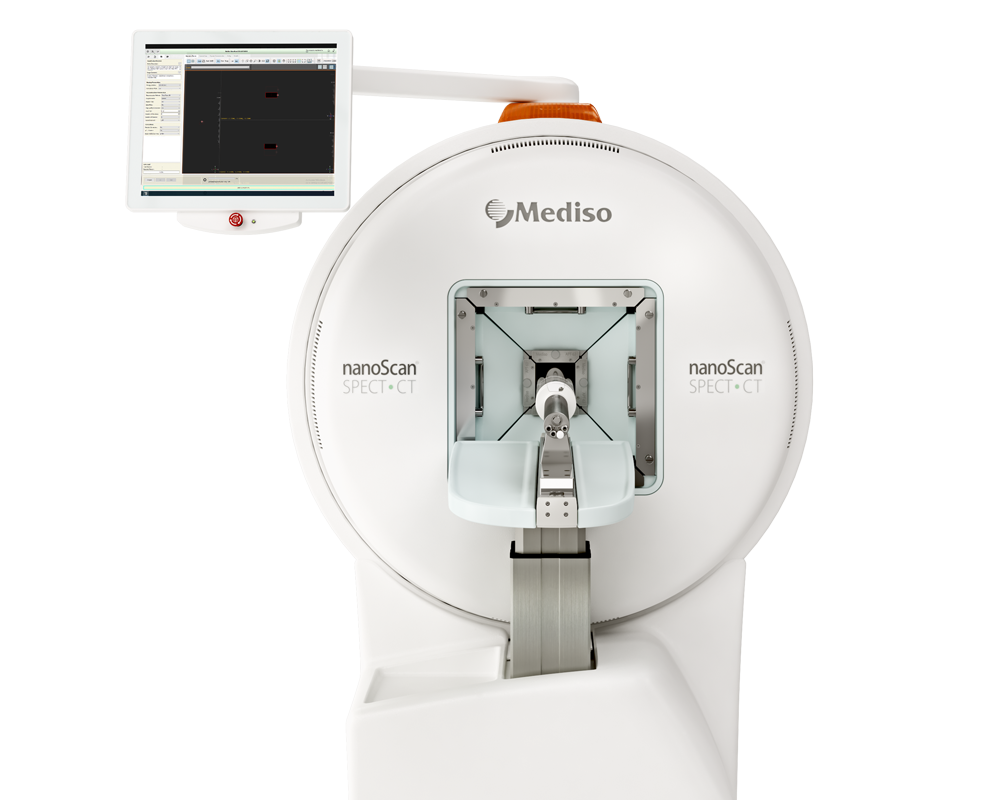
Image data were acquired using a small animal, dual detector nanoSPECT/CT system (Mediso, Hungary). Each detector consists of a 262 × 255 × 6.35 mm thallium-doped sodium-iodide (NaI(Tl)) crystal, connected with a hexagonally-arranged photomultiplier tube array. A 20% energy window over each photopeak of interest was used for data collection, with a single photopeak centred at 140 keV for 99mTc (t1/2 = 6 h), and two photopeaks centred at 48 and 75 keV for 161Tb (t1/2 = 7 d).
Anaesthesia was induced and maintained using 5 and 2% isoflurane, respectively, in a 1 L/minute oxygen flow. Mice were cannulated (tail vein) and transferred to the imaging cell, where temperature and respiration were monitored throughout scanning. Animals were centred in the FOV under a single pinhole collimator with aid of an x-ray scout scan. Once positioned, 60-second planar scintigraphy frames were repeatedly acquired over 1 h. The first acquisition was synchronised with radiotracer administration. Animals were injected (200 µL) with either [99mTc]TcO4− (18.5 ± 2.7 MBq, n = 11), [99mTc]TcO4−BAB cage (18.0 ± 3.6 MBq, n = 11), or [161Tb]Tb-PSMA-617 (22.7 MBq, n = 1). For animals that underwent SPECT/CT immediately after the dynamic planar scintigraphy (n = 3 [99mTc]TcO4−, n = 3 [99mTc]TcO4−BAB cage and n = 1 [161Tb]Tb-PSMA-617), the single pinhole collimator was exchanged for a multi-pinhole collimator and a 30 min whole-body SPECT/CT scan was acquired. Syringes and catheters containing radiotracers were measured before and after injection to calculate the injected activity.
Whole-body quantitative accuracy of 1-minute frames acquired by planar scintigraphy between 5 and 60 min post-injection was 101.1 ± 6.6% and 97.3 ± 8.2% for [99mTc]TcO4− (n = 11) and [99mTc]TcO4−BAB cage (n = 11), respectively. The same data quantified without sensitivity correction led to a considerable underestimation in whole-body activity, returning 76.6 ± 7.6% and 73.3 ± 6.9% for the same radiopharmaceuticals, respectively (Fig. 3).
Planar scintigraphy images show uptake in thyroid and stomach in animals injected with [99mTc]TcO4− and uptake in thyroid, stomach and liver for animals injected with [99mTc]TcO4−BAB cage (Fig. 4).
[99mTc]TcO4− (n = 11) scintigraphy at 1 h post-injection showed stomach and thyroid uptake of 45.7 ± 11.1%IA and 6.1 ± 3.4%IA, respectively (Fig. 5A). For the subset of animals (n = 3) which underwent dynamic planar scintigraphy followed by SPECT/CT, the stomach and thyroid activities returned by planar scintigraphy at the 1-hour timepoint were 50.2 ± 16.4 and 4.4 ± 0.4%IA and were 48.3 ± 10.2 and 4.7 ± 1.0%IA by SPECT, respectively (Fig. 5B). Decay-corrected whole-body activities represented 101.5 ± 3.8% (SPECT) and 104.9 ± 11.2% (planar scintigraphy) of the recorded injected activities.
[99mTc]TcO4−BAB cage (n = 11) scintigraphy at 1-hour post-injection returned stomach, thyroid and liver values of 29.6 ± 9.2%IA, 3.0 ± 1.1%IA and 22.3 ± 4.0%IA, respectively (Fig. 5C). For the subset of animals (n = 3) which underwent dynamic planar scintigraphy followed by SPECT/CT, uptake observed at the 1-hour timepoint of planar scintigraphy was 25.5 ± 3.1%IA (stomach), 4.1 ± 1.6%IA (thyroid) and 21.0 ± 4.2 (liver) %IA. Similarly, uptake observed in SPECT images was 23.1 ± 3.3%IA (stomach), 3.9 ± 2.3%IA (thyroid) and 14.8 ± 1.9 (liver) %IA at 1-hour post-injection (Fig. 5D). Decay-corrected whole-body activities represented 96.7 ± 6.7% (SPECT) and 94.1 ± 8.4% (planar scintigraphy) of the calculated injected activities.
For [161Tb]Tb-PSMA-617 (n = 1), increase in tumour uptake was observed over time, with values of 5.2%IA (planar) and 5.3%IA (SPECT) at 1 h post-injection (Fig. 6). The whole-body activity obtained by SPECT was 105.2% of the recorded injected activity. Whole-body quantitative accuracy of 1-minute frames acquired by planar scintigraphy between 5- and 60-minutes post-injection was 94.6 ± 3.6%. The same data quantified without sensitivity correction underestimated whole-body activity with 76.6 ± 3.1% quantitative accuracy.
- In the presented work, the ability to acquire quantitative whole-body dynamic planar scintigraphy images on a small animal SPECT/CT scanner has been demonstrated and incorporated into a SPECT imaging workflow with 99mTc- and 161Tb-based radiopharmaceuticals.
- Sensitivity deteriorates away from the centre of the FOV when acquiring projections using single pinhole collimators and requires sensitivity-based corrections to return quantitative data and avoid underestimations.
- Whole-body and organ-specific activities can be accurately determined by planar scintigraphy, and adoption of such methods in preclinical SPECT studies adds value in radiopharmaceutical development and dosimetry measurements.
How can we help you?
Don't hesitate to contact us for technical information or to find out more about our products and services.
Get in touch