Preclinical evaluation of an antibody-based companion diagnostic for CD44v6 expressing cancer
2026.04.01.
Maryam Oroujeni et al., Nuclear Medicine Communications, 2026
Abstract
Development of companion diagnostics for targeted radionuclide therapy is critical, especially for full-size antibodies with prolonged circulation times. Engineering antibodies to modify their in-vivo pharmacokinetics, such as incorporating neonatal Fc receptor (FcRn)-binding mutations, can potentially enable earlier imaging timing and improved patient stratification. This study aimed to evaluate the impact of FcRn-binding mutations on the in-vitro binding characteristics and in-vivo biodistribution and imaging performance of a CD44v6-targeting full-size antibody, UU-40, labeled with different radionuclides, and to assess its potential as a companion diagnostic.
The study involved engineering UU-40 with LALA and IAHA mutations, evaluating specific binding, internalization, and affinity using in-vitro cell assays. Biodistribution and imaging studies [PET and single-photon emission computed tomography (SPECT)] were conducted in mice carrying human tumor xenografts in a dual-nuclide setting.
The FcRn mutations (LALA/IAHA) did not affect antibody specificity or affinity, which was target-specific and affinity remained in the subnanomolar range. Biodistribution studies demonstrated that the residualizing radiometal label (177Lu) resulted in higher liver and spleen uptake compared with the nonresidualizing 125I-label, leading to reduced tumor-to-organ ratios. Tumor uptake was higher in A431 xenografts, with peak accumulation at 24 h postinjection. SPECT and PET imaging confirmed superior contrast at later time points (~24 h) with 125I-UU-40LALA/IAHA, while earlier imaging with 68Ga was hindered by increased nonspecific accumulation.
FcRn-binding mutations in full-size antibodies significantly alter their in-vivo pharmacokinetics without affecting binding affinity or specificity. Introducing these mutations enables earlier imaging time points, enhancing the potential for companion diagnostics in clinical settings.
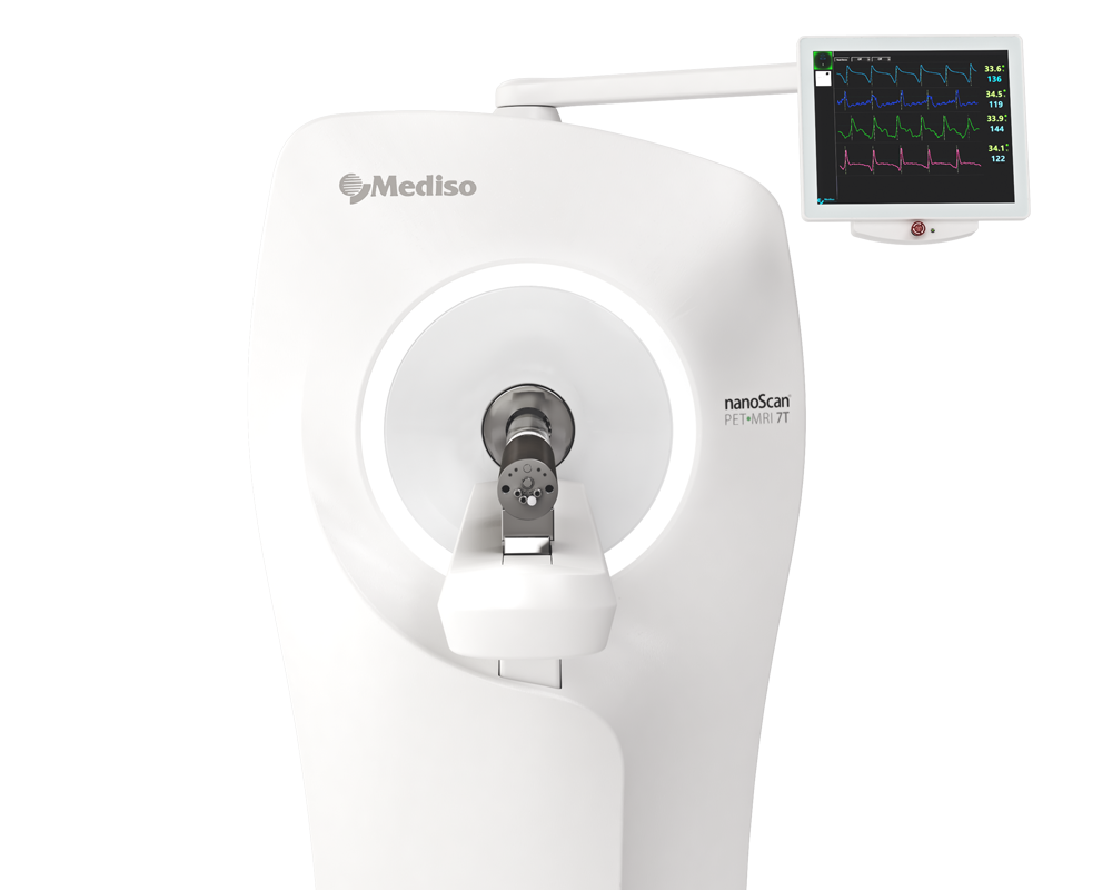
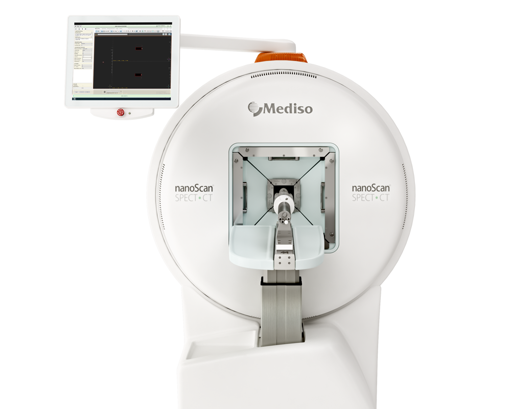
Results from nanoScan® SPECT/CT and nanoScan® PET/MRI 3T
For PET-computed tomography (CT), imaging subjects were anesthetized using sevoflurane (3–4% in 50%/50% medical oxygen/air at 450 ml/min). First, a whole-body CT scan was acquired with the following parameters: scan method: semi-circle FOV; projections 480; X-ray, 50 kVp and 610 μA; binning, 1 : 4; acquisition time, 4 min. A PET scan was performed on the same scan range as the CT, for 60 min. Subjects were placed on a heated bed to prevent hypothermia. The PET raw data were reconstructed in Nucline software, Budapest, Hungary (3.04.015.000) using the TeraTomo 3D algorithm with six subsets, four iterations, and corrected for scattering and attenuation artifacts. The CT raw files were reconstructed using Filter Back Projection. PET and CT Dicom files were co-registered, and analyzed using PMOD v 4.105 (PMOD Technologies Ltd., Fällanden, Switzerland).
For SPECT-CT, subjects were anesthetized using sevoflurane (3–4% in 50%/50% medical oxygen/air at 450 ml/min). First, a whole-body CT scan was acquired with the following parameters: scan method: semi-circle FOV; projections 480; X-ray, 50 kVp and 610 μA; binning, 1 : 4; acquisition time, 4 min. A SPECT scan was performed on the same scan range as the CT, for 30 min with the following parameters: frame time, 15 s; acquisition over energy windows of 125I (28.4 keV). Subjects were placed on the heated bed to prevent hypothermia. The SPECT raw data were reconstructed in Nucline software (3.04.015.000) using the TeraTomo 3D algorithm with three subsets, nine iterations, and corrected for scattering and attenuation artifacts. The CT raw files were reconstructed using Filter Back Projection. SPECT and CT Dicom files were co-registered, and analyzed using PMOD v 4.105 (PMOD Technologies Ltd., Switzerland).
One mouse carrying two large A431 xenografts (one on each posterior flank) was injected with a dual-nuclide solution containing 68Ga-UU-40LALA/IAHA (3.5 MBq) and 125I-UU-40LALA/IAHA (9.1 MBq). The PET/CT demonstrated specific tumor uptake of the 68Ga-labeled radioconjugate at 4 h p.i., albeit with relatively poor contrast (Fig. 4a). Comparatively greater uptake was demonstrated in the SPECT/CT at 5 h p.i. using 125I-UU-40LALA/IAHA (Fig. 4b). The highest uptake with low levels of background signal was demonstrated at 24 h p.i. using the 125I-UU-40LALA/IAHA (Fig. 4c). High signals were detected in the thyroid and urinary bladder in the SPECT scan performed at 5 h p.i. due to free 125I and the images were blocked during image analysis. Similarly, the thyroid was blocked in the SPECT scan performed at 24 h p.i.
Fig 4.: Small-animal PET/CT and SPECT/CT of a mouse carrying dual A431 xenografts. The animal was injected with dual-nuclide solution containing 68Ga (3.5 MBq) and 125I-UU-40LALA/IAHA (9.1 MBq). (a) PET-imaging at 4 h using 68Ga-UU-40LALA/IAHA. (b) SPECT-imaging at 5 h p.i. of 125I-UU-40LALA/IAHA. (c) SPECT-imaging at 24 h p.i. of 125I-UU-40LALA/IAHA. The same mouse was imaged at all time points (N = 1).

Full article on journals.lww.com
How can we help you?
Don't hesitate to contact us for technical information or to find out more about our products and services.
Get in touch