Toward Bifunctional Chelators for Thallium-201 for Use in Nuclear Medicine
2022.07.08.
Alex Rigby et al, Bioconjugate chemistry, 2022
Summary
Auger electron therapy exploits the cytotoxicity of low-energy electrons emitted during radioactive decay that travel very short distances (typically <1 μm). 201Tl, with a half-life of 73 h, emits ∼37 Auger and other secondary electrons per decay and can be tracked in vivo as its gamma emissions enable SPECT imaging. Despite the useful nuclear properties of 201Tl, satisfactory bifunctional chelators to incorporate it into bioconjugates for molecular targeting have not been developed. H4pypa, H5decapa, H4neunpa-NH2, and H4noneunpa are multidentate N- and O-donor chelators that have previously been shown to have high affinity for 111In, 177Lu, and 89Zr. Herein, we report the synthesis and serum stability of [nat/201Tl]Tl3+ complexes with H4pypa, H5decapa, H4neunpa-NH2, and H4noneunpa. All ligands quickly and efficiently formed complexes with [201Tl]Tl3+ that gave simple single-peak radiochromatograms and showed greatly improved serum stability compared to DOTA and DTPA. [natTl]Tl-pypa was further characterized using nuclear magnetic resonance spectroscopy (NMR), mass spectroscopy (MS), and X-ray crystallography, showing evidence of the proton-dependent presence of a nine-coordinate complex and an eight-coordinate complex with a pendant carboxylic acid group. A prostate-specific membrane antigen (PSMA)-targeting bioconjugate of H4pypa was synthesized and radiolabeled. The uptake of [201Tl]Tl-pypa-PSMA in DU145 PSMA-positive and PSMA-negative prostate cancer cells was evaluated in vitro and showed evidence of bioreductive release of 201Tl and cellular uptake characteristic of unchelated [201Tl]TlCl. SPECT/CT imaging was used to probe the in vivo biodistribution and stability of [201Tl]Tl-pypa-PSMA. In healthy animals, [201Tl]Tl-pypa-PSMA did not show the myocardial uptake that is characteristic of unchelated 201Tl. In mice bearing DU145 PSMA-positive and PSMA-negative prostate cancer xenografts, the uptake of [201Tl]Tl-pypa-PSMA in DU145 PSMA-positive tumors was higher than that in DU145 PSMA-negative tumors but insufficient for useful tumor targeting. We conclude that H4pypa and related ligands represent an advance compared to conventional radiometal chelators such as DOTA and DTPA for Tl3+ chelation but do not resist dissociation for long periods in the biological environment due to vulnerability to reduction of Tl3+ and subsequent release of Tl+. However, this is the first report describing the incorporation of [201Tl]Tl3+ into a chelator-peptide bioconjugate and represents a significant advance in the field of 201Tl-based radiopharmaceuticals. The design of the next generation of chelators must include features to mitigate this susceptibility to bioreduction, which does not arise for other trivalent heavy radiometals.
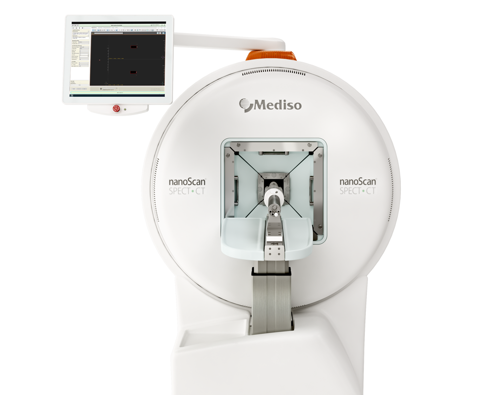
Results from nanoScan® SPECT/CT
Healthy SCID/beige animals (male 5-7 weeks old, n = 3 per radiotracer) were injected via tail vein injection under isoflurane anesthesia (1.5-2.5% in oxygen at 1 L/min) with [201Tl]TlCl (17-22.9 MBq), [201Tl]TlCl3 (11.2-23.8 MBq), or [201Tl]Tl-pypa-PSMA (14.1-16.9 MBq). Mice were then
kept under continuous anesthesia on a heated pad for the duration of the experiment (1 h), and one mouse per group was imaged by SPECT/CT until 1 h post injection.
To study tracer uptake in tumors, SCID/beige mice (male 5-7 weeks old, n = 3 per group) were injected subcutaneously with DU145-PSMA or DU145 cells (4 × 10⁶ cells in 100 mL
PBS) in the left shoulder. Once tumors had reached 5-10 mm in diameter (4-5 weeks after inoculation), [201Tl]Tl-pypaPSMA (10.7-24.5 MBq, 20 mmol) was administered via tail vein injection under isoflurane anesthesia. Mice were maintained under continuous anesthesia and imaged by
SPECT/CT for up to 2 h post injection. SPECT images were reconstructed using the HiSPECT (Scivis GmbH) reconstruction software package at 0.3 mm isotropic voxel size using standard reconstruction with 35% smoothing and nine iterations. After euthanasia, organs were harvested from the mice, weighed, and gamma counted. Images were analyzed using VivoQuant 2.5, enabling the delineation of regions of interest (ROIs) for quantifcation of radioactivity. ROIs for the tumor and organs (heart, muscle, etc.) were drawn using CT images, and volumes were determined. The total activity in the whole animal (excluding the majority of tail, out of SPECT field of view) at the time of [201Tl] agents’ administration was defned as the injected activity (IA), and the percentage of injected activity per cm3 (% IA/cm3) and amount of radioactivity in tissues (MBq) were determined. A 5 mL syringe with 3 mL of [201Tl]TlCl (40 MBq) was used to calibrate the SPECT/CT and ensure correct co-registration between the SPECT and CT.
SPECT/CT images showed that compared to [201Tl]Tl-pypa-PSMA, 201Tl administered as either Tl+ or Tl3+ has an initially high heart uptake at 15 min (4.5% and 3.6% IA (percentage injected activity), respectively) followed by washout, a high degree of retention in the kidneys (10.0 - 12.9% IA), and relatively low excretion via the urine/bladder (<1.7% IA at all time points) (Figure 6A). In contrast,
[201Tl]Tl-pypa-PSMA showed a lower myocardial accumulation at 15 min (2.1% IA) and signifcant [201Tl]Tl activity associated with the urine/bladder (8.4% at 60 min). Ex vivo biodistribution data showed that blood values were low for [201Tl]TlCl, [201Tl]TlCl3, and [201Tl]Tl-pypa-PSMA
with only 0.24, 0.18, and 0.19% activity, respectively, present in blood at 1 h post injection (p.i.) (Figure 6C). [201Tl]TlCl and [201Tl]TlCl3 have a high heart uptake of 10.3 ± 0.1% injected
activity per gram (IA/g) and 15.4 ± 2.6% IA/g at 1 h p.i., respectively, while [201Tl]Tl-pypa-PSMA showed a lower uptake (8.0 ± 0.4% IA/g) (Figure 6C), consistent with SPECT imaging analysis. All three 201Tl compounds were predominantly cleared via the kidneys, with [201Tl]TlCl having
74.4 ± 6.3% IA/g, [201Tl]TlCl3 having 104.5 ± 6.9% IA/g, and [201Tl]Tl-pypa-PSMA having 61.0 ± 3.0% IA/g accumulating in kidneys at 1 h p.i. Clearance through the liver was much lower for all three groups, with [201Tl]TlCl having 12.3 ± 0.6% IA/g, [201Tl]TlCl3 having 17.5 ± 2.0% IA/g, and [201Tl]Tl-pypa-PSMA having 15.3 ± 4.2% IA/g accumulating in the liver by 1 h p.i.

Figure 6. (A) In vivo images of [201Tl]TlCl, [201Tl]TlCl3, and [201Tl]Tl-pypa-PSMA at 15, 30, 45, and 60 min in healthy animals (n = 3 per group). (B) Regions of interest (ROIs) drawn from the SPECT images around organs of interest (bladder, heart, and kidneys) for [201Tl]TlCl, [201Tl]TlCl3, and [201Tl]Tl-pypa-PSMA at 15, 30, 45, and 60 min (n = 1 per radiotracer). (C) The ex vivo biodistribution of [201Tl]TlCl, [201Tl]TlCl3, and [201Tl]Tl-pypa-PSMA in healthy SCID beige mice culled at 1 h post injection (n = 3 per radiotracer).
The biodistribution of [201Tl]Tl-pypa-PSMA was studied in SCID/beige mice bearing either (i) DU145 PSMAexpressing tumors (PSMA-positive) or (ii) DU145 tumors that do not express the PSMA receptor (PSMA-negative) to determine if [201Tl]Tl-pypa-PSMA accumulated in prostate cancer tissues via PSMA receptor binding. This model has previously been used to show the PSMA-specifc uptake of
tracers. Each group of mice was administered [201Tl]Tl-pypaPSMA (10.7-24.5 MBq, 20 nmol) prior to SPECT/CT scanning for 2 h. At the conclusion of the SPECT/CT scan, each mouse was culled, and organs were dissected, weighed, and counted for radioactivity to obtain quantitative data on
radiotracer biodistribution. SPECT imaging analysis indicated that radioactivity
concentration in DU145 PSMA-positive tumors was consistently higher than in DU145 PSMA-negative tumors and, at early time points only, this difference was statistically
signifcant. At 30 min, the 201Tl radioactivity concentration in PSMA-positive DU145 tumors measured 3.5 ± 1.4% IA/g (p = 0.0219) and decreased to 2.9 ± 0.9% IA/g at 2 h p.i. (Figure 7C). For PSMA-negative DU145 tumors, 201Tl radioactivity concentration at 30 min was 2.1 ± 0.2% IA/g and remained steady until 2 h p.i. Biodistribution data 2 h p.i. corroborated SPECT imaging analysis: 201Tl concentration at 2 h p.i. in DU145 PSMA-positive tumors measured 3.7 ± 2.8% IA/g, and in the PSMA-negative tumors, this 201Tl radioactivity concentration measured 2.9 ± 1.5% IA/g (Figure 7B). Imaging
and ex vivo biodistribution data further evidenced that [201Tl]Tl-pypa-PSMA is cleared from the blood mainly via a renal pathway, with high levels of radioactivity observed in the
kidneys and bladder/urine evident in both imaging and ex vivo biodistribution data.
Ex vivo biodistribution data also indicated that the tumor/ blood ratio for PSMA-positive tumors (11.1 ± 1.4) was signifcantly higher than that for PSMA-negative tumors (3.9 ± 3.0) at 2 h p.i. (p = 0.0385). The tumor/muscle ratio was similarly higher in mice bearing PSMA-positive tumors (ratio
of 1.5 ± 0.4) than in mice bearing PSMA-negative tumors (ratio of 0.7 ± 0.2) (Figure 7E). SPECT image analysis was also used to determine tumor/muscle ratios for [201Tl]Tl-pypa-PSMA. The tumor/muscle ratio for animals bearing PSMAnegative tumors was approximately 1 from 30 min to 2 h p.i. However, the tumor/muscle ratio for animals bearing PSMApositive tumors measured 2.1 ± 0.7 at 30 min and decreased to 1.2 ± 0.4 at 2 h p.i.

Figure 7. (A) In vivo SPECT image (0−30 min) of [201Tl]Tl-pypa-PSMA in mice bearing DU145 positive and negative tumors at 0−30 min. SG = salivary glands, T = tumor, L = liver, K = kidneys, and B = bladder. (B) Ex vivo biodistribution of [201Tl]Tl-pypa-PSMA in mice bearing DU145 positive and negative tumors 2 h p.i. (n = 3 per group). (C) Uptake in DU145 PSMA-positive and PSMA-negative tumors using regions of interest drawn from the SPECT images at 30, 60, 90, and 120 min. Tumor to blood (D) and muscle (E) ratios were calculated using biodistribution data (2 h p.i.). Tumor to blood ratios were taken from ROIs drawn on the SPECT images at various time points (F).
Full article on pubs.acs.org
Wie können wir Ihnen behilflich sein?
Bitte kontaktieren Sie uns für technische Informationen und Unterstützung jeglicher Art in Zusammenhang mit unseren Entwicklungen und Produkten.
Kontaktformular
