Nuclear imaging-guided PD-L1 blockade therapy increases effectiveness of cancer immunotherapy
2020.11.08.
Hannan Gao et al., Journal of ImmunoTherapy of Cancer, 2020
Summary
The interaction between programmed death receptor-1 (PD-1) and its ligand (PD-L1) inhibits the function of effector T cells and the priming of naive T cells, leading to impaired antitumor immunity. Therefore the blockade of PD-1/PD-L1 signaling pathway has been a breakthrough in cancer therapy, but the response rate in solid tumors is only 20-30%. In this study, a new radiolabeled nanobody-based imaging probe 99mTc-MY1523 targeting PD-L1 was developed for the enhanced therapeutic efficacy of PD-L1 blockade immunotherapy.
Results show that the new probe has high binding affinity and specificity to PD-L1. SPECT/CT imaging revealed fast blood clearance, renal-route excretion and satisfactory tumor uptake.
As the timing for PD-L1 blockade therapy is crucial due to dynamic and heterogeneous expression of PD-L1 in tumors, it was essential to prove that SPECT/CT imaging is able to detect changes in the PD-L1 expression. Therefore PD-L1 expression was increased by interferon-γ (IFN-γ) treatment of tumor bearing mice and PD-L1 expression was determined by SPECT/CT imaging and verified by the flow cytometry. It proves that SPECT/CT imaging of 99mTc-MY1523 can be used to monitor PD-L1 expression in tumors in a real time, dynamic and quantitative manner.
The PD-L1 blockade therapy initiated during the therapeutic time window determined by 99mTc-MY1523 SPECT/CT imaging significantly enhanced the therapeutic efficacy: the tumor growth was dramatically suppressed, and the survival time of mice was evidently prolonged.
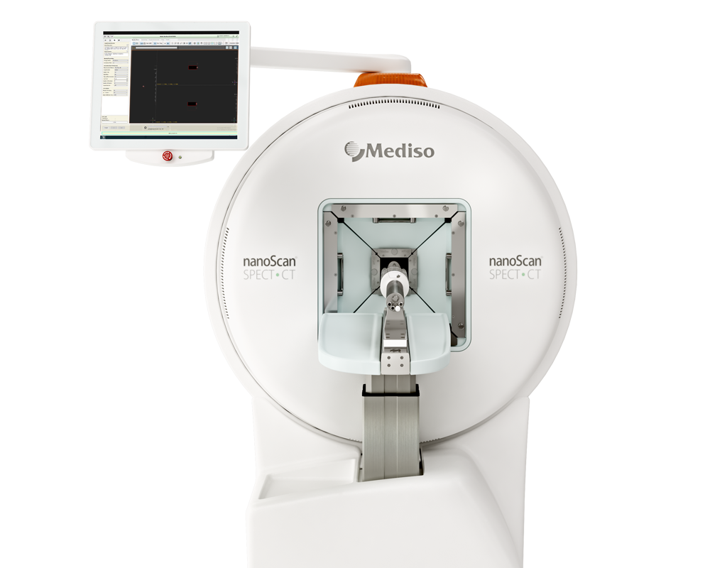
Results from nanoScan SPECT/CT
Three types of tumor cells (4T1, A20 or MC-38) were inoculated subcutaneously into the right flank of BALB/c or C57/BL6 mice, respectively. Four days later they were injected i.t. with PBS or IFN-γ for 5 days, and then were subjected to SPECT/CT imaging.
Mice were injected intraveneously with 18 MBq 99mTc-MY1523 and imaged at 2 hours p.i. (n=4) using the nanoScan SPECT/CT system with the following parameters: pinhole SPECT (peak: 140 keV, 20% width; frame time: 25 s), helical CT (50 kVp, 0.67 mA, rotation 210°, exposure time: 300 ms). SPECT and CT images were merged using the Nucline software V.2.0 (Mediso Ltd.). The regions of interest were drawn for the determination of tumor sizes (mm3) and radioactivity (Bq), then the tumor uptake was calculated as percentage injected dose per volume (%ID/cc).
- Results show increased tumor uptake of 99mTc-MY1523 compared to the corresponding control group in all animal models:

When imaging results showed the upregulated PD-L1 expression in tumors after IFN-γ intervention on day 8 and 12 after tumor cell inoculation, the mice were subjected to PD-L1 blockade therapy: they were ip. injected with 200μg αPD-L1 antibody twice with 4 days interval, while using PBS, IFN-γ and αPD-L1 antibody without IFN-γ intervention as controls. Tumor sizes were measured twice a week and calculated as volumes (mm3)=length×width×height/2.
- As shown on the figure below, although IFN-γ intervention expedited the tumor growth, the imaging-guided therapy dramatically improved the therapeutic efficacy. The tumor growth was significantly suppressed, and three of five tumors completely disappeared. Compared to control groups, the survival time of mice in the treated group was also remarkably prolonged.

Wie können wir Ihnen behilflich sein?
Bitte kontaktieren Sie uns für technische Informationen und Unterstützung jeglicher Art in Zusammenhang mit unseren Entwicklungen und Produkten.
Kontaktformular