Dose predictions for [177Lu]Lu-DOTA-EGF F(ab′)2 in NRG mice with HNSCC patient-derived tumour xenografts based on [64Cu]Cu-DOTA-EGF F(ab′)2 – implications for a PET theranostic strategy
2021.08.12.
Anthony Ku, Raymond M. Reilly et al., EJNMMI Radiopharmacy and Chemistry
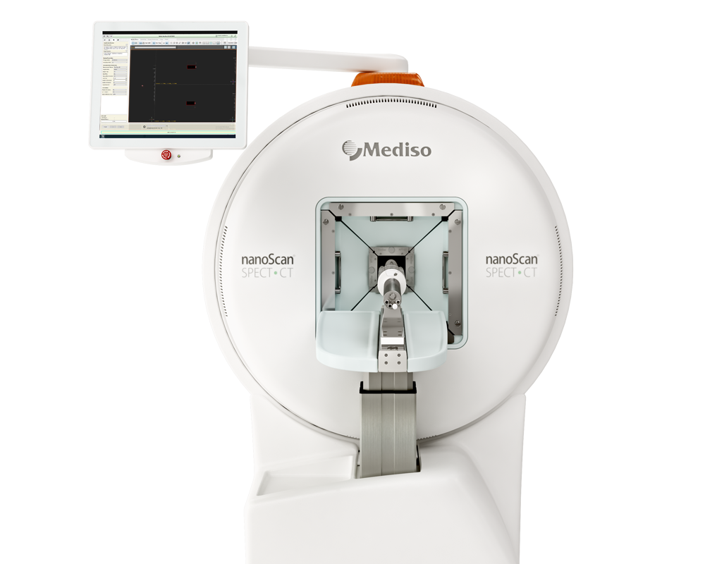
nanoScan® SPECT/CT/PET triple modality system was used to follow a PET and a SPECT radiotracer biodistribution and predict radiation equivalent doses for [177Lu]Lu-DOTA-panitumumab F(ab′)2 radioimmunotherapy.
Background
Epidermal growth factor receptors (EGFR) are overexpressed on many head and neck squamous cell carcinoma (HNSCC). Radioimmunotherapy (RIT) with F(ab')2 of the anti-EGFR monoclonal antibody panitumumab labeled with the βparticle emitter, 177Lu may be a promising treatment for HNSCC. The aim was to assess the feasibility of a theranostic strategy that combines positron emission tomography (PET) with [64Cu]Cu-DOTA-panitumumab F(ab')2 to image HNSCC and predict the radiation equivalent doses to the tumour and normal organs from RIT with [177Lu]Lu-DOTA-panitumumab F(ab')2.
Results from nanoScan® SPECT/CT/PET
- Researchers reported for the first time radiation equivalent dose predictions for [177Lu]Lu-DOTA-panitumumab F(ab´)2 based on biodistribution studies and ROI analysis of microPET/CT images in NRG mice with HNSCC patient-derived xenografts tumours after i.v. administration of [64Cu]Cu-DOTA-panitumumab F(ab´)2.
- Biodistribution (BOD) studies at 6, 24 or 48 h post-injection (p.i.) of [64Cu]Cu-DOTA-panitumumab F(ab')2 (5.5–14.0 MBq; 50 μg) or [177Lu]Lu-DOTA-panitumumab F(ab')2 (6.5 MBq; 50 μg) in NRG mice with s.c. HNSCC patient-derived xenografts (PDX) overall showed no significant differences in tumour uptake but modest differences in normal organ uptake were noted at certain time points.
- [64Cu]Cu-DOTA-panitumumab F(ab')2 and [177Lu]Lu-DOTA-panitumumab F(ab')2 tumour uptake were significantly higher (P < 0.05) than a non anti-EGFR antibody [177Lu]Lu-DOTA-trastuzumab F(ab')2 uptake, demonstrating EGFR-mediated tumour uptake
- Human doses from administration of [177Lu]Lu-DOTA-panitumumab F(ab')2 predicted that a 2 cm diameter HNSCC tumour in a patient would receive 1.1–1.5 mSv/MBq and the whole body dose would be 0.15–0.22 mSv/MBq.

Fig. 3 Posterior whole-body coronal microPET/CT images of a NRG mouse bearing s.c. implanted HNSCC PDX at a. 6 h, b. 24 h or c. 48 h p.i. of [64Cu]Cu-DOTA-panitumumab F(ab´)2. The tumour is shown by the white arrow. At 6 h p.i., the mediastinum (blood pool; white arrowhead), liver (blue arrowhead) and kidneys (broken red circles) are visualized, while at 24 and 48 h p.i., the liver was the only normal organ visualized.

Fig. 4 Posterior whole-body coronal microSPECT/CT images of a NRG mouse bearing s.c. implanted HNSCC PDX at a. 6 h, b. 24 h or c. 48 h p.i. of [177Lu]Lu-DOTA-panitumumab F(ab´)2 or d. 24 h p.i. of irrelevant [177Lu]Lu-DOTA-trastuzumab F(ab´)2. The tumour is shown by the white arrow. At 6 h p.i., the mediastinum (blood pool; white arrowhead), liver (blue arrowhead) and kidneys (broken white circles) were visualized, while at 24 and 48 h p.i., the liver and kidneys were the main normal organs visualized.
Full article published at EJNMMI Radiopharmacy and Chemistry
Wie können wir Ihnen behilflich sein?
Bitte kontaktieren Sie uns für technische Informationen und Unterstützung jeglicher Art in Zusammenhang mit unseren Entwicklungen und Produkten.
Kontaktformular

