Beyond the microcirculation: sequestration of infected red blood cells and reduced flow in large draining veins in experimental cerebral malaria
2024.03.16.
Anja M. Oelschlegel et al., Nature Communications, 2024
Summary
Cerebral malaria (CM) is a severe neurological complication of human malaria mostly caused by the parasite Plasmodium falciparum (Pf). It is the most common non-traumatic encephalopathy in the world with a high mortality rate. Globally, an estimated number of 608,000 patients died of malaria in 2022, while 96% of the malaria-related deaths occurred in sub-Saharan Africa, primarily in infants and children below the age of 5 years (WHO, 2023).
Patients with acute infection can present with a diffuse CM encephalopathy, a rapid progressive coma, and seizures without return to consciousness. Magnetic resonance imaging (MRI) findings in children in sub-Saharan Africa indicate that increased brain volume and brain swelling are hallmarks of the disease, and patients die from brain herniation. MRI measures of brain edema strongly correlate with the outcome in pediatric CM.
The key pathological findings in post-mortem human brains are intravascular sequestration of and congestion with infected red blood cells (iRBCs). In contrast to other parasitic diseases of the CNS, Pf-infected red blood cells in CM remain primarily intravascular. However, in murine experimental CM (ECM) some iRBCs have also been observed perivascularly in the parenchyma. The cascade of events linking infection and brain swelling has remained elusive and is much debated. Theories focus either on a cytokine storm triggering inflammation and affecting vascular integrity or mechanical obstructions of cerebral blood flow (CBF) due to iRBC sequestration in the microcirculation. The former is primarily based on ECM models, while the latter originates from post-mortemhistology of CM patients. While the relevance of ECM to CM continues to be viewed differently, there is growing consensus that studies with experimental models of CM are central to better define pathogenesis and identify early signatures that distinguish disease progression from parasite propagation. One central controversy is to which extent iRBC sequestration in cerebral microcapillaries, a distinctive hallmark of CM, also contributes to the disease in ECM. Therefore, insights into the temporal development of disease events in combination with their regional distribution might provide a better understanding of the pathophysiological processes leading to CM.
As the spatial patterns of brain swelling differ in different subgroups of patients, it could be highly informative to correlate at, or close to, the onset of cerebral edema, the spatial patterns of iRBC sequestration with spatial patterns of edema development and potential perfusion deficits, which are to be expected upon mechanical obstruction. However, such studies are hardly feasible in humans, especially in the most affected countries with limited access to the imaging technologies needed.
Herein, the authors address these questions in murine ECM focusing on Plasmodium berghei ANKA (PbA) infected C57BL/6 wild-type (wt) mice. They extend parts of our study to murine malaria models that do not develop ECM or brain pathology. After studying brain iRBC accumulation in infected mice using quantitative real-time-PCR (q-PCR), the authors use single-photon emission computed tomography (SPECT) for imaging in vivo the distribution of 99mTechnetium (99mTc)-labeled iRBCs in the brain at day 5 post-infection (p.i.), an early stage of the disease when first clinical symptoms start appearing. They combine this approach with T2-weighted (T2w) MRI for edema monitoring as well as SPECT-imaging of CBF and MR-angiography. These imaging approaches allow us to detect potential sequelae of iRBC sequestration on cerebral perfusion at different spatial levels, i.e., capillary flow and large draining veins. The authors also relate these data to histological data at different time points after infection and to the expression of inflammation markers, as studied by q-PCR and immunohistochemistry, with and without anti-malarial treatment.
Results from the nanoScan SPECT/CT
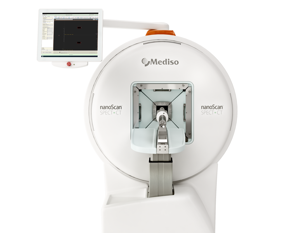
SPECT/CT imaging was performed with a four-head NanoSPECT/CTTM scanner (Mediso, Hungary). Animals were scanned under gas anesthesia (1.2–1.5% isoflurane, 850 ml/min O2). CT and SPECT were co-registered. Head scans were accompanied by two co-registered CT scans, one before and one after the SPECT scan, to control for motion artefacts (none detected). CT scans were made at 45 kVp, 177 μA, with 180 projections, 500 ms per projection, and 96 μm isotropic spatial resolution, reconstructed with the manufacturer´s software (InVivoScope 1.43) at isotropic voxel-sizes of 200 μm for whole-body scans and 100 µm for head scans. SPECT scans were made using nine-pinhole mouse brain apertures with 1.2 mm pinhole diameters providing a nominal spatial resolution ≤1 mm. Photopeaks were set to the default values of the NanoSPECT/CT for 99mTc (140 keV ± 5%). SPECT images were reconstructed using the manufacturer´s software (HiSPECTTM, SCIVIS, Germany) at an isotropic voxel size of 448 μm in whole-body scans and 300 µm in head scans.
Fig 1. B–F shows the spatial distribution of the differences in 99mTc-labeled iRBC contents between mice at day 5 p.i. and day 0 in C57BL/6 wt mice, G–I C57BL/6 prf−/− mice and J, K BALB/c mice. All color scales are in units of SUV. All SPECT images are differences of group-mean d5 p.i. minus d0 data. B Overlay of a midsagittal section of the SPECT-difference image on a group-mean MR-angiography from d0 mice. C Top view on a volume rendered difference SPECT overlaid on the reference MR. D, G, J Midsagittal sections of the difference SPECT in the different groups. E, F, H, I, K For precise localization of the peak regions, the SPECT-difference data sets were overlaid on an anatomical reference MR. The MR dataset is from an ex vivo measurement76 and includes a signal-intense rostral rhinal vein (gray arrows in C, F, and I). Note the precision of the alignment of the group-mean CTs (blue) and the MR (F, I). Peak differences in labeled iRBCs are found at the rostral (arrows pointing right downward) and the caudal confluences of the sinuses (arrows pointing left downward). Note also increased accumulation in midsagittal sinus (asterisk in B–D), straight sinus (arrowhead in B, D), and transverse sinus (arrowheads in C). L Boxplot diagram (min, max. median, 1st and 3rd quartil) of accumulation of 99mTc-labeled iRBCs in rrv and confluence of the sinuses (cs) VOIs in d5 p.i. relative to d0 mice (C57BL/6 wt d0 n = 7, d5 n = 8; C57BL/6 prf−/− and BALB/c n = 6, 9 weeks old female mice in all groups). The VOIs are shown in the sagittal section in (E) (white squares). *p < 0.05, **p < 0.01, ***p < 0.001, two-way ANOVA in (A), two-tailed unpaired heteroscedastic t-test in (L).
- The authors show early stage iRBC sequestration and reduced flow in large draining veins only in P. berghei-infected brains that are on a trajectory to ECM.
- They propose that in CM and ECM pathology edema and hemorrhage formation are causally linked to iRBC-induced reductions in venous efflux in large draining veins downstream of the microcirculation and postcapillary venules.
Wie können wir Ihnen behilflich sein?
Bitte kontaktieren Sie uns für technische Informationen und Unterstützung jeglicher Art in Zusammenhang mit unseren Entwicklungen und Produkten.
Kontaktformular