Willkommen bei Mediso

Bitte wählen Sie Ihre Region aus:

North America
Europa

Bitte wählen Sie Ihre Sprache:

Auswählen

Aldehydes alter TGF-β signaling and induce obesity and cancer

2024.09.24.

Xiaochun Yang,.., Scott K Lyons, Joseph R Merrill,.., Lopa Mishra, et al.

Cell reports, 2024

Summary


Obesity and fatty liver diseases—metabolic dysfunction-associated steatotic liver disease (MASLD) and metabolic dysfunction-associated steatohepatitis (MASH)—affect over one-third of the global population and are exacerbated in individuals with reduced functional aldehyde dehydrogenase 2 (ALDH2), observed in approximately 560 million people. Current treatment to prevent disease progression to cancer remains inadequate, requiring innovative approaches. We observe that Aldh2−/− and Aldh2−/−Sptbn1+/− mice develop phenotypes of human metabolic syndrome (MetS) and MASH with accumulation of endogenous aldehydes such as 4-hydroxynonenal (4-HNE). Mechanistic studies demonstrate aberrant transforming growth factor β (TGF-β) signaling through 4-HNE modification of the SMAD3 adaptor SPTBN1 (β2-spectrin) to pro-fibrotic and pro-oncogenic phenotypes, which is restored to normal SMAD3 signaling by targeting SPTBN1 with small interfering RNA (siRNA). Significantly, therapeutic inhibition of SPTBN1 blocks MASH and fibrosis in a human model and, additionally, improves glucose handling in Aldh2−/− and Aldh2−/−Sptbn1+/− mice. This study identifies SPTBN1 as a critical regulator of the functional phenotype of toxic aldehyde-induced MASH and a potential therapeutic target.

Results from the nanoScan PET/CT

  • Aldh2−/−Sptbn1+/− (ASKO) mice had significantly elevated visceral fat (epididymal white adipose tissue [eWAT]) accumulation (Figures 1D and S2B) on a normal chow (NC) diet
  • ASKO mice on the NC diet showed a trend toward a greater fat volume (31.57% ± 2.81% vs. 18.97% ± 2.93%) and less lean tissue volume (47.73% ± 1.45% vs. 56.13% ± 3.03%) than WT mice (Figure 1 E and 1F)
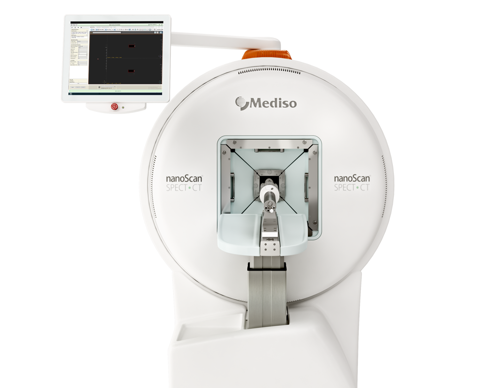
To assess the volume of body fat and lean tissue, mice were scanned by microCT. Anesthesia was induced with 3% isoflurane in oxygen and maintained at 2% isoflurane. Whole-body CT was acquired on a Mediso nanoScan PET/CT (Mediso USA, Arlington, VA, USA), calibrated to Hounsfield Units. 720 projections were acquired at 50 kVp energy and 189 μm. As exposure, and images were reconstructed in Nucline software (Mediso USA) using a filtered back-projection algorithm with a Blackman filter to a voxel size of 188 μm isotropic.
 
CT images were analyzed in VivoQuant 2021 (Invicro, Needham, MA, USA) using a custom script (available upon request). Briefly, after removing the imaging cradle from the CT image, a lower threshold of −500 HU was applied to segment the whole mouse and calculate its volume. Next, the lungs were segmented and combined with an automatically-thresholded airspace region of interest. This region of interest (ROI) was dilated to cover regions between dense tissue and airspace, which would otherwise be falsely counted as less-dense fat. Finally, a threshold between −400 and −100 HU was applied to the whole-body image, not including the dilated airspace ROI, to segment fat, and a threshold between −100 and 300 HU was applied to the whole-body image, excluding the airspace and urinary bladder, to segment lean tissue. The volumes of these fat and lean ROI’s were then divided by the whole-body volume to calculate the percent fat and lean tissue values.
 
Figure 1. C-F
(C) Body weight of the four genotypes was measured from 3 to 12 months of age. Statistically significant differences in body weight were determined by one-way ANOVA with Bonferroni’s multiple comparisons test comparing the averaged body weight of each mutant over time to the WT (n = 4–9). Data are presented as mean ± SEM. p < 0.05.
(D) Epididymal white adipose (eWAT) tissue weight of the indicated mice genotypes at 44–48 weeks old. Data are presented as mean ± SEM. Significant differences in eWAT weight were determined by pairwise t tests (n = 7–10 mice/genotype). ∗∗p < 0.01.
(E) Lean tissue volume and fat volume were measured by whole-body CT scan of each genotype on normal diet. Representative cross-sectional images (at L5) of each genotype are shown. Light blue (arrowheads) indicates lean tissue, and dark blue (arrows) indicates fat tissue.
(F) Graph showing the percentage of fat volume, lean tissue volume, and lean tissue/fat ratio (n = 2 mice/genotype). Data are presented as mean ± SEM.
 
 
Figure S2 A-D
(A) Expression of ALDH2 and SPTBN1 proteins in the liver of mice of indicated genotypes (WT, Sptbn1+/-, Aldh2-/- and ASKO) by Western Blot.
(B) Representative images of the visceral fat and whole-body fat/lean mass by CT scan of indicated genotypes at age of 44-48 weeks on normal diet.
(C) Representative cross image (at L5) of each genotype on Western diet, light blue (Arrow Heads) indicates lean mass, dark blue (Arrows) indicate fat mass.
(D) Graph of fat volume, lean tissue volume, and lean tissue/fat ratio (n=2 mice/genotype), *p<0.05. It shows increased fat volume, decreased lean tissue volume and a reduced lean tissue/fat ratio in ASKO mice on Western diet.
 

Wie können wir Ihnen behilflich sein?

Bitte kontaktieren Sie uns für technische Informationen und Unterstützung jeglicher Art in Zusammenhang mit unseren Entwicklungen und Produkten.

Kontaktformular