Promising potential of [177Lu]Lu-DOTA-folate to enhance tumor response to immunotherapy—a preclinical study using a syngeneic breast cancer model
2020.10.19.
Patrycja Guzik et al, European Journal of Nuclear Medicine and Molecular Imaging, 2020
Summary
Purpose It was previously demonstrated that radiation effects can enhance the therapy outcome of immune checkpoint inhibitors. In this study, a syngeneic breast tumor mouse model was used to investigate the effect of [177Lu]Lu-DOTA-folate as an immune stimulus to enhance anti-CTLA-4 immunotherapy.
Methods In vitro and in vivo studies were performed to characterize NF9006 breast tumor cells with regard to folate receptor (FR) expression and the possibility of tumor targeting using [177Lu]Lu-DOTA-folate and nanoScan SPECT/CT. A preclinical therapy study was performed over 70 days with NF9006 tumor-bearing mice that received vehicle only (group A); [177Lu]Lu-DOTA-folate (5 MBq; 3.5 Gy absorbed tumor dose; group B); anti-CTLA-4 antibody (3 × 200 μg; group C), or both agents (group D). The mice were monitored regarding tumor growth over time and signs indicating adverse events of the treatment.
Results [177Lu]Lu-DOTA-folate bound specifically to NF9006 tumor cells and tissue in vitro and accumulated in NF9006 tumors in vivo. The treatment with [177Lu]Lu-DOTA-folate or an anti-CTLA-4 antibody had only a minor effect on NF9006 tumor growth and did not substantially increase the median survival time of mice (23 day and 19 days, respectively) as compared with untreated controls (12 days). [177Lu]Lu-DOTA-folate sensitized, however, the tumors to anti-CTLA-4 immunotherapy, which became obvious by reduced tumor growth and, hence, a significantly improved median survival time of mice (> 70 days). No obvious signs of adverse effects were observed in treated mice as compared with untreated controls.
Conclusion Application of [177Lu]Lu-DOTA-folate had a positive effect on the therapy outcome of anti-CTLA-4 immunotherapy. The results of this study may open new perspectives for future clinical translation of folate radioconjugates.
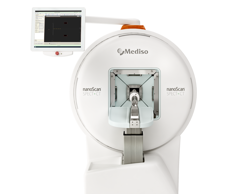
Results from nanoScan SPECT/CT
Six-seven weeks old female FVB/NCrl mice were fed with a folate-deficient rodent diet. After acclimatization for 5–7 days, they were were subcutaneously inoculated with 2.5 × 10⁶ NF9006 tumor cells in 100 μL PBS. 12–14 days after NF9006 tumor cell inoculation (tumor volume: ~ 100–300 mm3) CT scans of 7.5 min duration time were followed by a SPECT scan of ~ 40 min of NF9006 tumor-bearing mice at 4 h and 24 h after injection of [177Lu]Lu-DOTA-folate (25 MBq, 0.5 nmol, 100 μL).
Results show:
- SPECT/CT imaging studies confirmed in vitro result: high uptake of [177Lu]Lu-DOTA-folate in NF9006 tumors and in the kidneys. The uptake in lymph nodes of the neck and armpits appeared specific to this mouse strain rather than related to the tumor as demonstrated in control experiments, performed with FVB mice without tumors in which the same distribution pattern was observed
 Full article on link.springer.com
Wie können wir Ihnen behilflich sein?
Bitte kontaktieren Sie uns für technische Informationen und Unterstützung jeglicher Art in Zusammenhang mit unseren Entwicklungen und Produkten.
Kontaktformular
