Cerebellar microglial Metabolic–Inflammatory coupling drives network progression in temporal lobe epilepsy
2026.06.12.
Yuanxia Wu et al., Meta-Radiology, 2026
Summary
Epilepsy is among the most common and disabling chronic neurological disorders, characterized by recurrent seizures arising from abnormal, hypersynchronous neuronal discharges. Increasing evidence indicates that epilepsy is not merely a focal disorder but rather a condition involving dysfunction of distributed networks. Temporal lobe epilepsy (TLE) is increasingly recognized as a network disorder that extends beyond the hippocampus. Among the extra-hippocampal regions, the cerebellum has emerged as a remote yet functionally connected node showing consistent metabolic alterations. However, its mechanistic contribution to seizure propagation and disease progression remains largely unclear. The cerebellum is traditionally regarded as a motor control center, but has recently been recognized as an important subcortical hub in epileptic networks. Anatomical and electrophysiological studies demonstrate the cerebellum's extensive bidirectional connectivity with multiple cortical and limbic regions. This connectivity suggests that the cerebellum can influence seizure dynamics. The aim of this study was to elucidate the contribution of cerebellar microglial activation to aberrant glucose metabolism and to determine whether metabolic-inflammatory coupling within the cerebellum drives seizure generalization and cognitive decline in TLE.
Adult male Sprague-Dawley rats were examined. Temporal lobe epilepsy was induced using the lithium chloride-pilocarpine method. Status epilepticus (SE) was defined as continuous stage IV-V seizures according to the modified Racine scale. Diazepam was administered 60 min after the onset of SE. Thirty days after SE, the rats underwent stereotactic implantation of a microinfusion pump targeting the dentate nucleus for local drug delivery. Minocycline or the c-Fos inhibitor T-5224 was infused for 14 days.
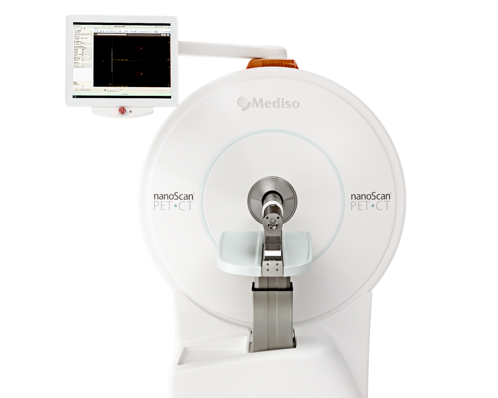
Rats underwent a longitudinal [18F]FDG microPET/CT scan at 1 day (acute phase), 14 days (latent phase) and 60 days (chronic phase) after SE using a nanoScan® PET/CT system (Mediso, Hungary). In the chronic phase, [18F]DPA-714 imaging was performed to assess microglial activation. The rats were fasted overnight, anesthetized and injected intravenously with ether [18F]FDG or [18F]DPA-714 through the tail vein. Scans were initiated 45 min or 30 min after injection respectively, with a 15-min static acquisition.
Bilateral cerebellar [18F]FDG SUVr increased significantly during the chronic phase but remained unchanged in the acute and latent stages. These data suggest that cerebellar hypermetabolism develops as a chronic adaptation during epilepsy progression.

Fig. 3. In vivo validation of cerebellar microglial activation in chronic-stage TLE rats using [18F]DPA-714 PET. (A) Representative coronal, sagittal, and axial [18F]DPA-714 PET/CT images acquired 60 days after status epilepticus (SE), demonstrating increased tracer uptake in the bilateral cerebellum. (B) Quantitative analysis of [18F]DPA-714 standardized uptake value ratios (SUVr), calculated using the pons as a reference region, showing significantly elevated cerebellar TSPO binding in TLE rats compared with controls.
Conclusion
These findings identify cerebellar metabolic-inflammatory coupling, driven by c-Fos-dependent microglial activation, as a key mechanism promoting epileptic network progression. Targeting this cerebellar axis provides a promising therapeutic avenue for precision neuromodulation in TLE.
How can we help you?
Don't hesitate to contact us for technical information or to find out more about our products and services.
Get in touch